![](https://viewpoint.online/wp-content/uploads/2020/09/P4-RE-colour-fundus-960x959.jpg 960w, https://viewpoint.online/wp-content/uploads/2020/09/P4-RE-colour-fundus-1280x1279.jpg 1280w, https://viewpoint.online/wp-content/uploads/2020/09/P4-RE-colour-fundus-300x300.jpg 300w, https://viewpoint.online/wp-content/uploads/2020/09/P4-RE-colour-fundus-160x160.jpg 160w, https://viewpoint.online/wp-content/uploads/2020/09/P4-RE-colour-fundus-480x480.jpg 480w, https://viewpoint.online/wp-content/uploads/2020/09/P4-RE-colour-fundus-640x640.jpg 640w, https://viewpoint.online/wp-content/uploads/2020/09/P4-RE-colour-fundus.jpg 1489w)
1 CET • 45 minutes read
OCT in glaucoma: case studies
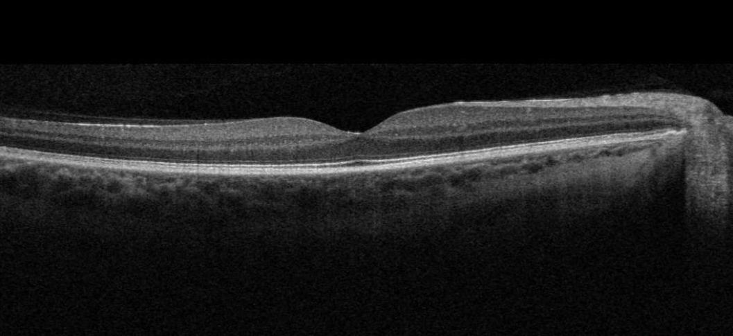
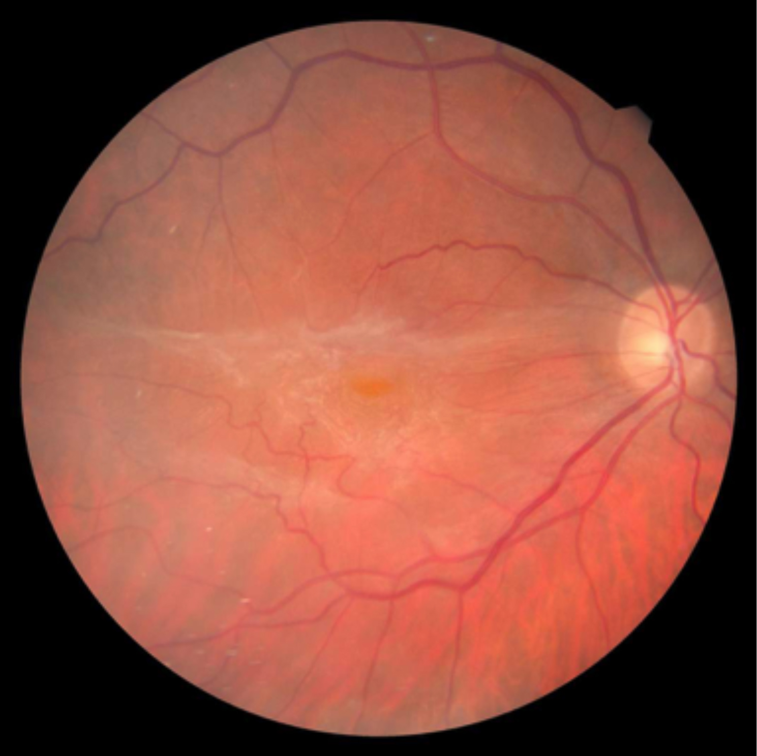
This series of case studies examines how clinicians can use OCT images to help in diagnosis and referral decision making in glaucoma.
What you will learn:
- Optometrists will have an enhanced understanding of the use of OCT for assessment of patients with or at risk of chronic open-angle glaucoma
- Optometrists will have an evidence-based understanding of the principles of use of OCT to assess various structures within the retina
- Optometrists will have an enhanced understanding of the identificatio