Non-exudative age-related macular degeneration

Referral priority: Low or moderate – depending on symptoms and clinical signs.

Cases with small-sized drusen should be monitored at local optometrist practice or referred to a local ophthalmologist if local guidelines require that.

Cases with confluent drusen and advanced non-exudative changes could be monitored by an experienced optometrist or referred to an ophthalmologist following local guidelines.

Click on one of the cards below to read more about the specific eye condition.

Written by
Marko Lukic
Edited by
Svein Tindlun and Jon Gjelle
Published
June 2023

Sections
01
Introduction

Go to section
02
Symptoms

Go to section
03
Clinical signs

Go to section
04
Diagnostic procedures

Go to section
05
Management and treatment

Go to section
06
References

Go to section
01

Introduction

Age-related macular degeneration (AMD) is a complex disease with genetic and environmental aetiology and is the leading cause of irreversible blindness.(1,2) AMD is the macula’s degenerative disease, characterized by a loss of visual acuity caused by degeneration of choriocapillaris, retinal pigment epithelium (RPE), and photoreceptors.(3,4)

The predicted number of patients diagnosed with AMD has been significantly growing, and the expected number of those diagnosed with AMD will be 288 million in 2040.(5)

The aetiology of AMD is multifactorial and involves an interplay of genetic, environmental, metabolic, and functional factors, including ageing, family history, smoking, high blood pressure, obesity, hypercholesterolemia, and arteriosclerosis.(6,7)

Understanding the correlation between risk factors rather than monitoring them individually is essential. It has been found that a higher BMI and a history of arterial hypertension, hypercholesterolemia, and atherosclerosis often occur together as risk factors for AMD.(8) There has been great progress in understanding the role of genes in the pathophysiology of the disease. Inflammation, or more specifically, an alternative complement pathway,  is at the centre of the pathomechanism of dry age-related macular degeneration. Treatment in the late phase of research is under development. However, a precise mechanism for the disease remains a challenge.

The hallmark of the disease is the presence of drusen.

Drusen are focal deposits of extracellular debris that typically form between the basal lamina of the RPE and the inner collagenous layer of Bruch’s membrane. Broadly speaking, the disease has two major subtypes: a non-exudative or so-called “dry type” and an exudative or so-called “wet type.”

The predicted number of patients diagnosed with AMD has been significantly growing, and the expected number of those diagnosed with AMD will be 288 million in 2040.

02

Symptoms

AMD is a slowly progressive condition, and patients at an early stage are usually asymptomatic. However, in some early cases, patients may complain of distorted vision (metamorphopsia), blurred vision, and central and paracentral scotoma. Patients usually complain they have problems with reading where some letters or a whole word is missing. As the disease progresses, the symptoms become more obvious. As the disease progresses, the symptoms become more obvious.

Patients with an exudative type of disease complain of a sudden decrease in vision. Remember – that patients who develop a more advanced stage of a non-exudative form of the disease (centre-involving geographic atrophy) may also complain of “sudden” deterioration in vision.

Patients with AMD have problems with central vision, whilst the peripheral vision remains intact (if there is no other concomitant eye condition like glaucoma). The loss of central visual acuity leads to a reduction in activities of daily living, as well as mobility problems and an increased risk of falls, fractures, and depression in older adults.(9)

Symptoms at a glance

Distorted vision (metamorphopsia)
Blurred vision
Central and paracentral scotoma
Problems with reading

03

Clinical signs

It is important to understand that the ageing process in the retina and macula is different from- age-related macular degeneration. Some similar changes make distinguishing normal ageing from pathological changes more difficult. For example, small drusen are often seen as an ageing process, while large and, more importantly, confluent drusen are pathological signs and considered part of the AMD.(10,11)

The AREDS Group constructed a classification system for the ARED Studies. It is especially useful in clinical practice.

Drusen

As has already been mentioned, the presence of drusen is the condition’s hallmark. Drusen can be described in – size, colour, morphology, and extent.(12,13) Drusen size is estimated by comparing the shortest diameter of a druse to the width of a significant – retinal vein as it crosses the edge of the optic disc. At this location, the retinal vein has an approximate diameter of 125 µm. Drusen are labelled small when they are less than half the width of the reference vein (<63 µm), medium, or intermediate when judged at least equal to half the vein width but less than the full width (63 µm to <125 µm), and large when they are at least as big as the full retinal vein width (≥125 µm).(14,15)

Drusen may be of white, pale yellow, or yellow colour. Regarding morphology, they are divided into hard and soft drusen. The soft drusen are medium or large-sized and have an amorphous appearance. Their borders are indistinct and may be confluent with one another. Some drusen may be calcified. Drusen may grow and coalesce or regress, resulting in atrophy and depigmentation.(16)

Classical drusen are located below the retinal pigment epithelium (RPE). However, small drusenoid deposits may be located above the RPE and are called subretinal drusenoid deposits or pseudodrusen.(17) Those deposits are also a risk factor for the development of late stages of AMD (geographic atrophy and type 3 CNV in exudative AMD).(18)

The AREDS Group constructed a classification system for – the AREDS studies. It is especially – useful in clinical practice:

  • No AMD – Presence of 0 to 5 small-sized drusen (<63 µm).
  • Early AMD – Multiple small-sized drusen, – a few moderate-size drusen (63 µm to <125 µm), or pigmentary changes.
  • Intermediate AMD – Extensive medium-sized drusen, at least one large drusen (≥125 µm), or extrafoveal geographic atrophy.
Image 1. Colour fundus photo of intermediate dry age-related macular degeneration; notice macular middle-sized and large-sized coalescent drusen with pigment migration.
Image 2. Colour fundus photo of intermediate dry age-related macular degeneration; notice large-sized drusen and extrafoveal patches of atrophy.
  • Advanced AMD – Fovea-involving geographic atrophy or signs of exudative age-related macular degeneration (subretinal or sub-RPE haemorrhage, serous fluid, serous PEDs, lipid exudates, fibrovascular scar).
Image 3. Advanced age-related macular degeneration; centre-involving atrophy and drusen.

Hyper- and hypo-pigmentary retinal changes

Focal hyperpigmentation areas are more common than hypopigmented areas. They represent clumps of pigment, which may be within RPE or intraretinally. Those changes may be located regardless of the position of the drusen. However, they are more often present at the border of drusen and drusenoid pigment epithelial detachments. Hypopigmented areas have – less pigment, and it has been postulated that hyperpigmentation may precede depigmentation.

Geographic atrophy

It is characterized by a local and sharp demarcation of atrophic outer retinal, RPE, and choriocapillaris layers.(19) Large choroidal vessels are seen at the area of atrophies. It starts in the perifoveal region and progresses towards the fovea. It is bilateral but, in most cases, asymmetrical. Likewise, it may represent a single or multifocal.

04

Diagnostic procedures

Amsler grid – a helpful tool to detect macular changes early. If the practice owns diagnostic imaging modalities like colour fundus photography and/or OCT, try to combine the results of the Amsler grid test and the images to better understand the test’s meaning. Remember, there may be false negative results!

Slit lamp examination – a straightforward approach to recognising the clinical features of the AMD on a slit lamp.

Colour fundus photography – a useful imaging modality to document the macular changes and monitor changes during follow-ups.

Optical coherence tomography – a golden standard imaging modality to recognize the macular changes related to AMD and to distinguish the entities which may look similar . Likewise, it is useful in recognizing drusen from pseudodrusen and – identifying signs of exudative AMD or geographic atrophy early.

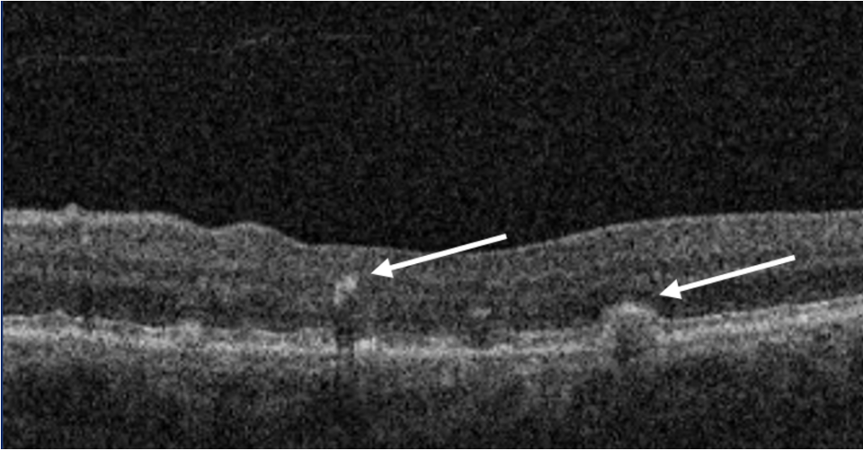
Image 4. OCT scan of dry AMD. The Blue arrow represents a druse. The white arrow represents pigment migration.
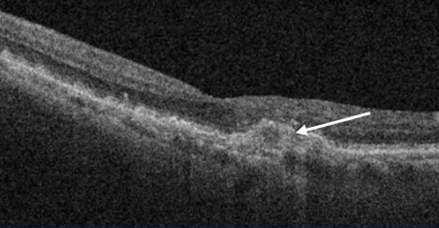
Image 5. OCT scan representing dry age-related macular degeneration. The yellow arrow represents coalescent drusen.
Image 6. Centre involving geographic atrophy. Notice missing RPE and increased shadow in the central macula due to atrophy.
05

Management and treatment

There is no current treatment to cure non-exudative age-related macular degeneration. However, the – AREDS studies brought evidence of the benefit of specific supplements, and the studies recommended two formulas.(20,21) Nowadays, AREDS 2 study formula is the one that should be recommended to patients with intermediate and more advanced cases of AMD.

Nutrient AREDS formula* AREDS 2 formula
Vitamin C 500 mg 500 mg
Vitamin E 400 IU 400 IU
Beta-carotene 15 mg
Copper (cupric oxide)** 2 mg 2 mg
Lutein 10 mg
Zeaxathin 2 mg
Zinc 80 mg 80 mg
* Not recommended for current or former smokers
** Added to avoid zinc-related copper deficiency
Mg = milligrams
IU = International units


Likewise, avoiding smoking is one of the most important recommendations you can suggest to your patients. Furthermore, increasing green vegetables on the weekly menu and wearing sunglasses are reasonable recommendations supported by some publications.(22,23)

Recent phases II and III trials have evaluated intravitreal C3 and C5 inhibitors and found that they were effective in reducing the growth of the lesion compared to sham injections.(24-26)


06

References

1 Friedman DS, O’Colmain BJ, Munoz B, Tomany SC, McCarty C, De Jong PT, Nemesure B, Mitchell P, Kempen J. Prevalence of age-related macular degeneration in the United States. Arch ophthalmic. 2004 Apr 1;122(4):564-72.

2 Sobrin L, Seddon JM. Nature and nurture-genes and environment-predict onset and progression of macular degeneration. Progress in retinal and eye research. 2014 May 1; 40:1-5.

3 Rosenfield PJ, Martidis A, Tennant M. Age-related macular degeneration. In: Yanoff M, Duker JS, Augsburger JJ, eds. Ophthalmology: Expert Consult. 3rd ed. Philadelphia, PA: Elsevier Mosby; 2009.

4 Shalev V, Sror M, Goldshtein I, Kokia E, Chodick G. Statin use and the risk of age-related macular degeneration in a large health organization in Israel. Ophthalmic epidemiology. 2011 Apr 1;18(2):83-90.

5 Wong WL, Su X, Li X, Cheung CM, Klein R, Cheng CY, Wong TY. Global prevalence of age-related macular degeneration and disease burden projection for 2020 and 2040: a systematic review and meta-analysis. The Lancet Global Health. 2014 Feb 1;2(2): e106-16.

6 Cheung LK, Eaton A. Age‐related macular degeneration. Pharmacotherapy: The Journal of Human Pharmacology and Drug Therapy. 2013 Aug;33(8):838-55.

7 Delcourt C, Souied E, Sanchez A, Bandello F. Development and validation of a risk score for age-related macular degeneration: the STARS Questionnaire. Investigative Ophthalmology & Visual Science. 2017 Dec 1;58(14):6399-407.

8 Bucan K, Lukic M, Bosnar D, Kopic A, Jukic T, Konjevoda S, Glavadanovic S, Gverovic Antunica A. Analysis of association of risk factors for age-related macular degeneration. European Journal of Ophthalmology. 2022 Jan;32(1):410-6.

9 Fong DS. Age-related macular degeneration: update for primary care. American Family Physician. 2000 May 15;61(10):3035-42.

10 Crabb JW, Miyagi M, Gu X, Shadrach K, West KA, Sakaguchi H, Kamei M, Hasan A, Yan L, Rayborn ME, Salomon RG. Drusen proteome analysis: an approach to the aetiology of age-related macular degeneration. Proceedings of the National Academy of Sciences. 2002 Nov 12;99(23):14682-7.

11 Bressler NM, Bressler SB, West SK, Fine SL, Taylor HR. The grading and prevalence of macular degeneration in Chesapeake Bay watermen. Archives of Ophthalmology. 1989 Jun 1;107(6):847-52.

12 Sarks JP, Sarks SH, Killingsworth MC. Evolution of soft drusen in age-related macular degeneration. Eye. 1994 May;8(3):269-83.

13 Klein R, Davis MD, Magli YL, Segal P, Klein BE, Hubbard L. The Wisconsin age-related maculopathy grading system. Ophthalmology. 1991 Jul 1;98(7):1128-34.

14 Ferris FL, Davis MD, Clemons TE, Lee LY, Chew EY, Lindblad AS, Milton RC, Bressler SB, Klein R. A simplified severity scale for age-related macular degeneration: AREDS Report No. 18. Archives of ophthalmology (Chicago, Ill.: 1960). 2005 Nov 1;123(11):1570-4.

15 Klein ML, Francis PJ, Ferris FL, Hamon SC, Clemons TE. Risk assessment model for development of advanced age-related macular degeneration. Archives of ophthalmology. 2011 Dec 1;129(12):1543-50.

16 Klein ML, Ferris III FL, Armstrong J, Hwang TS, Chew EY, Bressler SB, Chandra SR, AREDS Research Group. Retinal precursors and the development of geographic atrophy in age-related macular degeneration. Ophthalmology. 2008 Jun 1;115(6):1026-31.

17 Chen L, Messinger JD, Zhang Y, Spaide RF, Freund KB, Curcio CA. Subretinal drusenoid deposit in age-related macular degeneration: histologic insights into initiation, progression to atrophy, and imaging. Retina (Philadelphia, Pa.). 2020 Apr;40(4):618.

18 Spaide RF, Ooto S, Curcio CA. Subretinal drusenoid deposits AKA pseudodrusen. Survey of ophthalmology. 2018 Nov 1;63(6):782-815.

19 Fleckenstein M, Mitchell P, Freund KB, Sadda S, Holz FG, Brittain C, Henry EC, Ferrara D. The progression of geographic atrophy secondary to age-related macular degeneration. Ophthalmology. 2018 Mar 1;125(3):369-90.

20 Age-Related Eye Disease Study Research Group. A randomized, placebo-controlled, clinical trial of high-dose supplementation with vitamins C and E, beta carotene, and zinc for age-related macular degeneration and vision loss: AREDS report no. 8. Archives of ophthalmology. 2001 Oct 1;119(10):1417-36.

21 Chew EY, Clemons TE, SanGiovanni JP, Danis R, Ferris FL, Elman M, Antoszyk A, Ruby A, Orth D, Bressler S, Fish G. Lutein+ zeaxanthin and omega-3 fatty acids for age-related macular degeneration: the Age-Related Eye Disease Study 2 (AREDS2) randomized clinical trial. JAMA-Journal of the American Medical Association. 2013 May 15;309(19):2005-15.

22 Gopinath B, Liew G, Kifley A, Lewis JR, Bondonno C, Joachim N, Hodgson JM, Mitchell P. Association of dietary nitrate intake with the 15-year incidence of age-related macular degeneration. Journal of the Academy of Nutrition and Dietetics. 2018 Dec 1;118(12):2311-4.

23 Neelam K, Zhou SW, Eong KG. The role of blue light in the pathogenesis of age-related macular degeneration. Points de Vue. 2014(71):77.

24 https://investors.apellis.com/news-releases/news-release-details/apellis-completes-enrollment-two-phase-3-studies-targeted-c3

25 Jaffe GJ, Westby K, Csaky KG, Monés J, Pearlman JA, Patel SS, Joondeph BC, Randolph J, Masonson H, Rezaei KA. C5 inhibitor avacincaptad pegol for geographic atrophy due to age-related macular degeneration: a randomized pivotal phase 2/3 trial. Ophthalmology. 2021 Apr 1;128(4):576-86.

26 Liao DS, Grossi FV, El Mehdi D, Gerber MR, Brown DM, Heier JS, Wykoff CC, Singerman LJ, Abraham P, Grassmann F, Nuernberg P. Complement C3 inhibitor pegcetacoplan for geographic atrophy secondary to age-related macular degeneration: a randomized phase 2 trial. Ophthalmology. 2020 Feb 1;127(2):186-95.