Anterior uveitis

Referral priority: Moderate or urgent

Urgently refer all patients with hypertensive uveitis, bilateral uveitis, viral uveitis, and signs of more extensive disease to an ophthalmologist or a hospital following local guidelines. Likewise, an experienced eye care specialist should semi-urgently refer or treat all other forms of anterior uveitis (non-granulomatous, unilateral) while respecting local recommendations.

Click on one of the cards below to read more about the specific eye condition.

Written by
Marko Lukic
Edited by
Svein Tindlund and Jon Gjelle
Published
June 2023

Sections
01
Introduction

Read more
02
Symptoms

Read more
03
Clinical features

Read more
04
Diagnostic procedures

Read more
05
Management and treatment

Read more
06
References

Read more
01

Introduction

Uveitis is defined as inflammation of the uveal tract, which is further subdivided into anterior (iris and ciliary body) and posterior components (choroid). The inflammation may affect surrounding tissue like the retina and optic nerve.1 Uveitis, particularly posterior uveitis, is a common cause of preventable blindness, deemed a sight-threatening condition. It is responsible for between 10% and 15% of all cases of blindness and 30,000 new cases of legal blindness annually in the United States.(2,3) In Western countries, uveitis affects 200 per 100 000 people.(4) More than 35% of patients who develop the condition suffer severe visual impairment.(4,5)

The classification of uveitis is based on anatomical location and is as follows:

Type of uveitis  Primary location 
Anterior  Iris, ciliary body, or both 
Intermediate uveitis  Vitreous 
Posterior uveitis  Choroid 
Panuveitis  The entire uveal tract and vitreous 


Furthermore, uveitis is – additionally classified – by the following characteristics:(6)

  • Onset (sudden vs insidious)
  • Duration (limited, < 3 months in duration; persistent, >3 months in duration)
  • Course (acute, recurrent, or chronic)
  • Laterality (unilateral vs. bilateral)

The most common type of uveitis is acute anterior uveitis. According to the above classifications, uveitis specifically affects the anterior parts of the uveal tract and is characterised by a sudden onset and limited duration. The distribution of the condition is high, from 60.6% to 90.6%.(7) Most often, acute anterior uveitis is idiopathic (37.8%), which means it is not related to any illness or inflammation in the body.(7,8) However, genetic factors, trauma, diseases, and infections may trigger/cause anterior uveitis. The most common genetic relationship with acute anterior uveitis is the HLA-B27 genotype. Be aware that only 1% of patients who carry the HLA-B27 allele develop anterior uveitis.(9) The most associated conditions with anterior uveitis are seronegative arthropathies (ankylosing spondylitis, Reiter syndrome, psoriatic arthritis (no psoriasis per se), inflammatory bowel disease), juvenile rheumatoid arthritis (JRA), herpes virus, sarcoidosis, systemic lupus erythematosus (SLE), rheumatoid arthritis.

The pathomechanism of anterior uveitis is not completely clear. A leading theory is that exposure of an individual with a genetic predisposition to an infectious agent result- in cross-reactivity with ocular-specific antigens (molecular mimicry) with resultant iritis.(10) In clinical appearance, the anterior uveitis may be non-granulomatous or granulomatous.  Sometimes, based on clinical appearance, potential causes may be suspected (i.e., granulomatous AAU is present in Lyme disease, Tuberculosis, Syphilis, and herpes simplex virus).(11)

 

02

Symptoms

Typical symptoms in acute anterior uveitis are redness of the eye, pain, and photophobia (sensitivity to light). Vision may or may not be blurry. Acute anterior uveitis is usually a unilateral condition (mostly non-granulomatous inflammation), although different eyes may be affected at different times. The granulomatous kind of inflammation is more severe and usually bilateral.

Please note that patients may experience eye pain in different ways. Some may report no pain, while others may describe it as a foreign body sensation or an abrupt onset of a dull eye ache. The pain may be purely ocular or periorbital/temple pain.

Redness of the eye is not companied by discharge. However, photophobia (sunlight particularly) worsens eye discomfort.

Thoroughly obtaining a comprehensive medical history is crucial, involving the patient’s present and past personal history, surgical procedures, history of trauma, medications, and family medical history. Always ask about arthritis, rashes, shortness of breath, swollen lymph nodes, recent headaches, hearing difficulties, hair loss, pigment changes in the skin, a history of ocular trauma, recent insect bites, sexually transmitted diseases (STDs), TB exposure, blood in stools, and recent travel.(12,13,14)

03

Clinical features

Various clinical signs indicate the presence of intraocular inflammation. It is important to be familiar with them as the proper diagnosis is vital in uveitis conditions. It may be of sight-saving importance.

Intraocular pressure
Intraocular pressure is usually lower in the affected eye due to decreased production of aqueous secondary to an inflamed ciliary body. Sometimes, the IOP may be increased, and such uveitis is called hypertensive uveitis. It is commonly present in unilateral viral-related anterior uveitis.

Eye redness
Eye redness is present in the form of ciliary injection/flush. However, diffuse redness of conjunctiva may be present. Carefully examine conjunctiva for small nodules.

Keratitic precipitates
Keratitic precipitates are white blood cells present on the corneal endothelium. Small, white KPs are a sign of the non-granulomatous type of inflammation, while large, yellowish to brown KPs are significant for the granulomatous type of uveitis.

Image 1. Keratitic precipitates (non-granulomatous) attached to the inferior portion of corneal endothelium.

The presentation of corneal oedema and corneal decompensation may indicate viral aetiology.

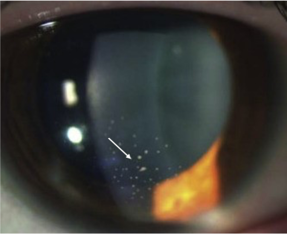
Competence in examining the anterior chamber is crucial in recognising and monitoring anterior uveitis. Flares and cells in the anterior chamber are signs of intraocular inflammation. Therefore, the cells in the AC should be graded by severity under high-magnification slit lamp examination in a 1 X 1-mm field of light, as described by The SUN Working Group Grading Scheme for Anterior Chamber Cells, as follows(15):

  • 0 < 1 cell
  • 0.5 = 1-5 cells
  • 1+ = 6-15 cells
  • 2+ = 16-25 cells
  • 3+ = 26-50 cells
  • 4+ = More than 50 cells

Flare is the presence of proteins in the anterior chamber, and the SUN Working Group proposed the below classification(15):

  • 0 = None
  • 1+ = Faint
  • 2+ = Moderate (iris and lens details clear)
  • 3+ = Marked (iris and lens details hazy)
  • 4+ = Intense (fibrin or plastic aqueous)

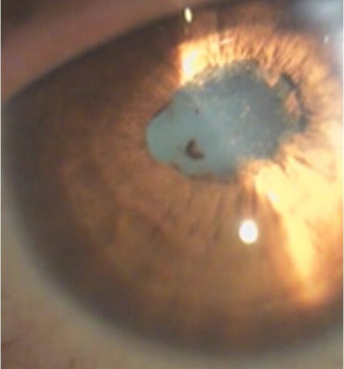
Examining the iris is very important in evaluating patients with uveitis. Chronic inflammation can develop anterior peripheral synechiae (adhesions between the iris and iridocorneal angle) or posterior synechiae (adhesions between the pupil margin and intraocular lens). Moreover, nodules on the iris may be observed, indicating inflammatory cell accumulation. These nodules are categorised into two types: Koeppe nodules, situated at the pupillary border, and Busacca nodules, located on the iris surface.


Image 2. Posterior synechiae present from 5-12 o'clock hours. Note the mild haziness of the photo. It is due to a moderate (2+ flare).

Patients with chronic uveitis may develop posterior subcapsular cataracts as a complication of the condition, taking steroids or both. Anterior vitreous cells are present in cases of cyclitis. Usually, the vitreous cells are signs of more extensive intraocular inflammation.

Image 3. Anterior chamber cells (orange arrows). To count anterior chamber cells, you need to use a high magnification, 1 × 1-mm slit beam through the anterior chamber and count the number of cells visible in the lit area.
04

Diagnostic procedures

Slit lamp examination is crucial in examining patients with anterior uveitis.

Colour fundus photos are useful in checking posterior eye segments and recognising signs of posterior uveitis.

Optical coherence tomography is a golden standard in diagnosing uveitis macular oedema. Anterior uveitis may be complicated with macular oedema, and OCT scans are essential in overall workup with uveitis patients.

05

Management and treatment

As highlighted in the section above, a thorough medical history is vital in evaluating uveitis patients.

During the first episode of patients with granulomatous inflammation, it is advisable to conduct laboratory and imaging workups. The recommended laboratory and imaging tests for such cases are as follows:(16)

  • Purified protein derivative (PPD) test or Quantiferon testing for tuberculosis.
  • A chest radiograph for sarcoidosis and TB should be obtained. In addition, a high-resolution chest CT should be considered if sarcoidosis is strongly suspected.
  • Sacroiliac, lumbar, or thoracolumbar spine radiographs may be ordered if ankylosing spondylitis is suspected.
  • Venereal Disease Research Laboratory (VDRL) test, FTA-ABS (or similar treponemal specific serology) test for syphilis.
  • CBC with differential.
  • Angiotensin-converting enzyme (ACE) test for sarcoidosis.
  • Lyme serology if Lyme disease is suspected.

An experienced eye care specialist should prescribe treatment of uveitis.
Topical corticosteroids are the mainstay of therapy and should be used aggressively during the initial phases of therapy. A long-acting cycloplegic agent (cyclopentolate) is used to relieve pain. However, it is recommended for the acute form of the condition rather than for chronic forms. Systemic immunosuppression therapy may be indicated for recurrent or chronic diseases and flares that are vision-threatening despite local therapy.

Remember that infectious causes primarily require treatment for the specific infectious agent.
Surgery (i.e., cataract surgery) is performed when the eye is quiescent for at least three months.

06

References

1 Dunn JP. Uveitis. Prim Care. 2015 Sep. 42 (3):305-23.

2 Nussenblatt RB. The natural history of uveitis. International ophthalmology. 1990 Oct;14(5):303-8.

3 DARRELL RW, WAGENER HP, KURLAND LT. Epidemiology of uveitis: incidence and prevalence in a small urban community. Archives of ophthalmology. 1962 Oct 1;68(4):502-14.

4 London NJ, Rathinam SR, Cunningham ET. The epidemiology of uveitis in developing countries. International Ophthalmology Clinics. 2010 Apr 1;50(2):1-7.

5 Rothova A, Suttorp-van Schulten MS, Frits Treffers W, et al. Causes and frequency of blindness in patients with intraocular inflammatory disease. Br J Ophthalmol. 1996;80:332–336.

6 Rao NA. Uveitis and other intraocular inflammations. Ophthalmology. 2nd ed. Philadelphia: Mosby. 2004:1105-238.

7 McCANNEL CA, Holland GN, Helm CJ, Cornell PJ, Winston JV, Rimmer TG, UCLA Community-Based Uveitis Study Group. Causes of uveitis in the general practice of ophthalmology. American Journal of Ophthalmology. 1996 Jan 1;121(1):35-46.

8 Rodriguez A, Calonge M, Pedroza-Seres M, Akova YA, Messmer EM, D’Amico DJ, Foster CS. Referral patterns of uveitis in a tertiary eye care center. Archives of ophthalmology. 1996 May 1;114(5):593-9.

9 Smith JR. HLA-B27–associated uveitis. Ophthalmol Clin North Am. 2002 Sep. 15(3):297-307.

10 https://eyewiki.aao.org/Acute_Anterior_Uveitis

11 Elnahry AG, Elnahry GA. Granulomatous Uveitis. InStatPearls [Internet] 2021 Apr 25. StatPearls Publishing.

12 Sanghvi C, Bell C, Woodhead M, Hardy C, Jones N. Presumed tuberculous uveitis: diagnosis, management, and outcome. Eye. 2011 Apr;25(4):475-80.

13 Kamal S, Kumar R, Kumar S, Goel R. Bilateral interstitial keratitis and granulomatous uveitis of tubercular origin. Eye & Contact Lens. 2014 Mar 1;40(2):e13-5.

14 Abderrahim K, Chebil A, Falfoul Y, Bouladi M, Matri LE. Granulomatous uveitis and reactive arthritis as manifestations of post-streptococcal syndrome. International ophthalmology. 2015 Oct;35(5):641-3.

15 Standardisation of Uveitis Nomenclature (SUN) Working Group. Standardisation of uveitis nomenclature for reporting clinical data. Results of the First International Workshop. American Journal of Ophthalmology. 2005 Sep 1;140(3):509-16.

16 https://emedicine.medscape.com/article/1209505-workup#c4