Bullous keratopathy

Referral priority: Moderate

All patients with bullous keratopathy should be referred to an ophthalmologist or a hospital following local guidelines.

Click on one of the cards below to read more about the specific eye condition.

Written by
Marko Lukic
Edited by
Svein Tindlund and Jon Gjelle
Published
June 2023

Sections
01
Introduction

Read more
02
Symptoms

Read more
03
Clinical features

Read more
04
Diagnostic procedures

Read more
05
Management and treatment

Read more
06
References

Read more
01

Introduction

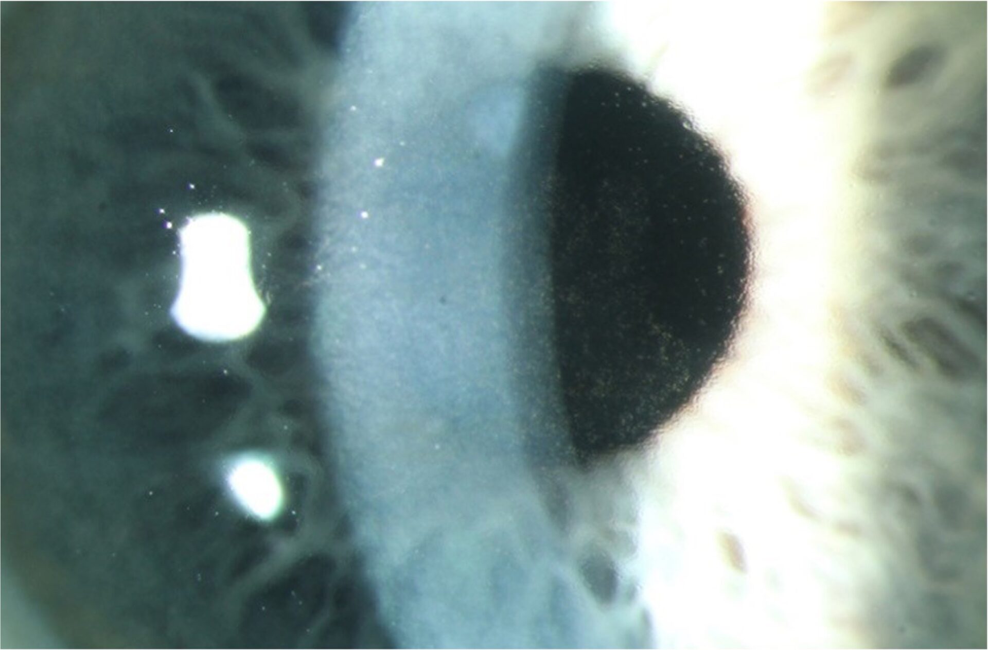
Bullous keratopathy refers to irreversible corneal oedema secondary to damage to the corneal endothelium. The most common cause is cataract surgery and intraocular (IOL) lens implantation; therefore, in literature is known as pseudophakic bullous keratopathy.(1,2) The bullous keratopathy may occur in aphakic eyes or in Fuchs’s corneal dystrophy.(3)

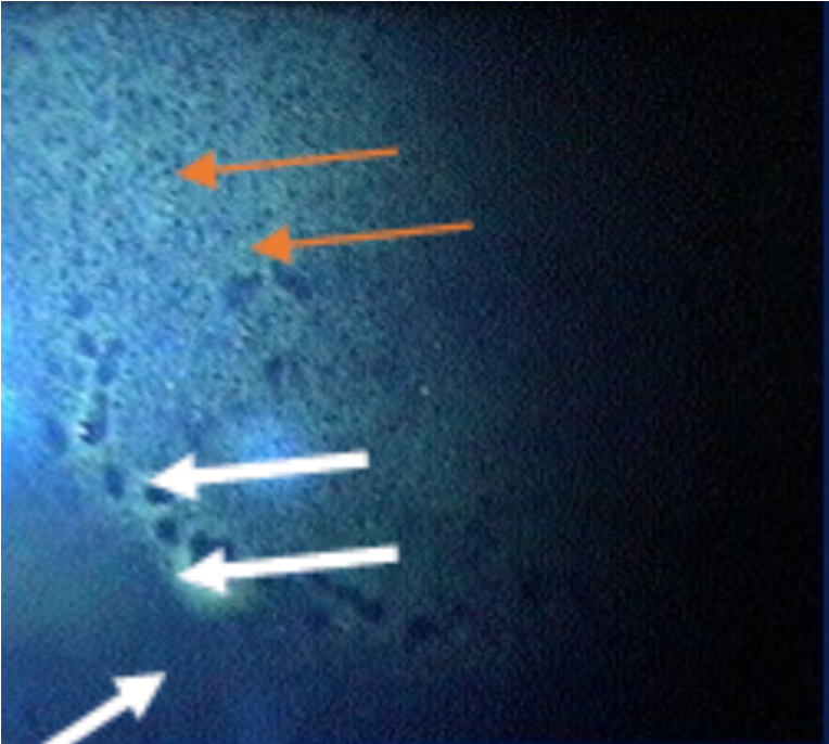
The cornea, to be transparent, needs to maintain a dehydrated state. The corneal endothelium is significant in keeping the cornea transparent, where endothelial cells work as a pump.(4) Any damage to the corneal endothelium brings a risk of developing corneal oedema and, consequently, bullous keratopathy. Firstly, the fluid enters the cornea’s stroma and then the epithelium. Epithelial oedema manifests with bullae.  Therefore, bullous keratopathy refers to the condition’s name. The endothelial cell density has decreased since birth, and the typical average cell density in an adult person is 2500-2700 cells/mm2. There must be a critically decreased cell density for bullous keratopathy to develop. The published studies revealed that bullous keratopathy occurs when the cell density is lower than 700 cells/mm2 or 300-500 cells/mm2.(1,5) It’s been thought that post, intra-, and post-operative cell loss contribute to the development of bullous keratopathy.(1)

Risk factors cause lower pre-operative endothelial cell density, like advanced age, pre-existing corneal Fuchs’s dystrophy, shallow anterior chamber, glaucoma, previous ocular trauma, diabetes, and chronic obstructive pulmonary disease.(6,7( Fuchs dystrophy is a genetic disorder that causes bilateral, progressive corneal endothelial cell loss, sometimes leading to symptomatic bullous keratopathy by age 50 to 60.(3)

It is estimated that 1-2% of people will develop persistent post-operative corneal oedema. Improving surgical techniques and the design of intraocular lenses improved the incidence of bullous keratopathy. Surgical trauma and pre-operative endothelial cell density are the main risk factors for developing the condition.(2)

02

Symptoms

In the early phase, patients may be asymptomatic. In more advanced stages, patients endorse gradual worsening of their vision. They have a positive history of cataract surgery on the affected eye. Usually, patients mention that the worst vision is in the morning, which has improved as the day progresses. The evaporation from the tear film is minimal at night with the eyes closed (then the tears are less hypertonic), and corneal oedema tends to be worse in the morning. Then, patients complain of halos of light, photophobia, foreign body sensation, and eye pain. Stromal oedema causes less light scattering and no pain. The pain is caused by the eruption of bullae located within the cornea’s epithelial layer.(8)

03

Clinical features

The physical examination plays a significant role in diagnosis.

Corneal oedema is defined as the increase in the thickness of the cornea due to the accumulation of extracellular fluid in epithelium and stroma, resulting in loss of corneal transparency. Corneal oedema results in increased corneal thickness.

Image 1. Presence of corneal oedema on a slit lamp (magnified image) in Fuchs’s corneal degeneration.

Descemet folds are secondary to damage to the corneal endothelial cells. They are not related only to bullous keratopathy but to all corneal conditions affecting endothelial cells’ health. The Descemet membrane is the basement membrane of endothelial cells.

Subepithelial bullae are bullous formations just below the corneal epithelium filled with fluid. They can rupture, which causes pain and corneal staining.

Image 2. Fluorescein staining of the cornea with bullous keratopathy. Bullae (white arrows) appear as negative staining. Small dots appearing as negative staining represent microcystic corneal oedema (orange arrows).

Stromal haze or opacification of the corneal stroma may also be present in bullous keratopathy. It is mostly a sign of a more chronic stage of the disease.

Corneal neovascularisations may also occur. They are not specific to the condition and happen in the upregulation of angiogenic cytokines.(9)

Image 3. Corneal scar with corneal neovascularisations. Both scar and corneal new vessels appear in different conditions at the end stage of the disease.
04

Diagnostic procedures

Corneal pachymetry is a measurement of corneal thickness. Patients with Fuchs endothelial dystrophy and a preoperative corneal thickness greater than 640 microns are at risk of developing the disease after cataract surgery.(11) Specular microscopy is a procedure that calculates endothelial cell density, shape, and size. Those with a density lower than 1000 cells/mm2 are at risk of developing bullous keratopathy. Then, patients with high variations in the size and shape of the endothelial cells, which indicate endothelial stress, have a chance of developing keratopathy.

05

Management and treatment

Early and mild corneal oedema can be treated medically with hypertonic eye drops and ointment (2-5% sodium chloride). These agents create a hypertonic tear film, drawing water out of the cornea. It is prudent to recommend that patients use the drops more frequently in the morning (4-5 times) and decrease frequency during the rest of the day (during the day, the natural tears become more hypertonic and help to reduce corneal oedema).

Patients with disrupted epithelial bullae are prescribed lubricant eye drops/ointments and local antibiotics to prevent infection.

Various surgical procedures can be used in the treatment of bullous keratopathy. Corneal transplantation is a definite treatment as it restores the function and anatomy of the corneal endothelium. Several keratoplasty procedures can be used; penetrating keratoplasty (PK), Descemet Membrane Endothelial Keratoplasty (DMEK), and Descemet Stripping Automated Endothelial Keratoplasty (DSAEK). DMEK has been shown to have better graft survival and lower rejection rates for cases of PBK when compared to PK or DSAEK.(10)

Amniotic membrane transplant (AMT), Anterior stromal puncture (ASP) and Phototherapeutic Keratectomy (PTK) can be treatments of choice, especially in pain control.

06

References

1 Narayanan R, Gaster RN, Kenney MC. Pseudophakic corneal oedema: a review of mechanisms and treatments. Cornea. 2006 Oct 1;25(9):993-1004.

2 Claesson M, Armitage WJ, Stenevi U. Corneal oedema after cataract surgery: predisposing factors and corneal graft outcome. Acta ophthalmologica. 2009 Mar;87(2):154-9.

3 https://www.merckmanuals.com/professional/eye-disorders/corneal-disorders/bullous-keratopathy

4 https://emedicine.medscape.com/article/1193218-overview#a5

5 Smolin G, Thoft RA, Dohlman CH. Endothelial function. The Cornea: Scientific Foundations and Clinical Practice. 3rd ed. Lippincott William & Wilkins: 1994. 635-643.

6 Ishikawa A. Risk factors for reduced corneal endothelial cell density before cataract surgery. Journal of Cataract & Refractive Surgery. 2002 Nov 1;28(11):1982-92.

7 Hasegawa Y, Nejima R, Mori Y, Sakisaka T, Minami K, Miyata K, Oshika T. Risk factors for corneal endothelial cell loss by cataract surgery in eyes with pseudoexfoliation syndrome. Clinical Ophthalmology (Auckland, NZ). 2016;10:1685.

8 Morishige N, Chikama TI, Yamada N, Takahashi N, Morita Y, Nishida T, Sonoda KH. Effect of pre-operative duration of stromal oedema in bullous keratopathy on early visual acuity after endothelial keratoplasty. Journal of Cataract & Refractive Surgery. 2012 Feb 1;38(2):303-8.

9 Feizi S, Azari AA, Safapour S. Therapeutic approaches for corneal neovascularization. Eye and Vision. 2017 Dec;4(1):1-0.

10 Tourtas T, Laaser K, Bachmann BO, Cursiefen C, Kruse FE. Descemet membrane endothelial keratoplasty versus descemet stripping automated endothelial keratoplasty. American Journal of Ophthalmology. 2012 Jun 1;153(6):1082-90.

11 Moshirfar M, Huynh R, Ellis JH. Cataract surgery and intraocular lens placement in patients with Fuchs corneal dystrophy: A review of the current literature. Current Opinion in Ophthalmology. 2022 Jan 1;33(1):21-7.