Central serous retinopathy and pachychoroidal conditions

Referral priority: Moderate or urgent – depending on clinical signs.

Urgently refer patients showing signs of secondary choroidal neovascularisation (CNV) to a hospital, following local guidelines. Refer patients who exhibit only central serous chorioretinopathy (CSR) features without (CNV) to an ophthalmologist for further management.

Click on one of the cards below to read more about the specific eye condition.

Written by
Marko Lukic
Edited by
Svein Tindlund and Jon Gjelle
Published
June 2023

Sections
01
Introduction

Read more
02
Symptoms

Read more
03
Clinical signs

Read more
04
Diagnostic procedures

Read more
05
Management and treatment

Read more
06
References

Read more
01

Introduction

Central serous chorioretinopathy (CSCR, CSC or CSR) is a disease characterised by localised and serous detachments of the neurosensory retina with or without focal pigment epithelial detachments (PEDs) and altered retinal pigment epithelium (RPE).(1) The condition is the 4th most common vision-threatening macular condition. It is a multifactorial and complex condition where leaking dilated choroidal vessels play a significant role.(3) It typically affects middle-aged men (72-88% of cases) who, in most cases, spontaneously recover within 3-4 months with an excellent visual prognosis.(4,5)

A group of patients may develop a chronic form of the disease, which has a worse visual prognosis.(1) Patients older than 50 have a higher risk of developing a bilateral and chronic form of the condition with a higher risk of developing secondary choroidal neovascular membrane (CNV). In addition, more female predominance is present in those older than 50.(6,7)

The pathomechanism of the condition is complex. Both endogenous and exogenous steroids have the most potent known association with CSR.(8,9) Increased choroidal vascular permeability is postulated to occur as a response to epinephrine-mediated vasospasm that is potentiated by steroids, leading to choroidal ischemia and vascular hyper-permeability.(10) Combined with RPE dysfunction, oncotic pressure in the choroidal space leads to fluid accumulation in the sub-retinal area.(11)

There are publications and evidence on other risk factors which may cause CSR clinical picture (Helicobacter pylori infection, sildenafil, ecstasy).(12,13,14) Furthermore, some chemotherapeutics, known as MEK/RAK inhibitors, and patients suffering from obstructive sleep apnoea can cause CSR.(15,16)

Nowadays, the knowledge of choroidal anatomy has increased, and CSR is considered part of the so-called pachychoroid spectrum of conditions (enlarged choroidal vessels). Other clinical entities where some clinical features may overlap are:(17)

  • Pachychoroid pigment epitheliopathy (PPE)
  • Pachychoroid neovasculopathy (PNV)
  • Polypoidal choroidal vasculopathy (IPCV) – is also considered part of AMD!
  • Focal choroidal excavation (FCE)
  • Peripapillary pachychoroid spectrum (PPS)
02

Symptoms

The symptoms depend on the location of the subretinal fluid. Remember that patients may be asymptomatic. The primary sign is a blurry central or paracentral vision with a relative scotoma. Patients may complain of metamorphopsia and micropsia (visual objects are perceived to be smaller than they are objectively sized). Likewise, patients may have reduced contrast sensitivity and colour saturation (dyschromatopsia).

Patients may have a hyperopic shift. Therefore, be aware of this when you check your refraction.

Take a detailed history and ask about the risk mentioned above factors. Despite the strong correlation between steroids and CSR, it seems that steroid eye drops do not increase the risk of CSR.

03

Clinical signs

Central serous retinopathy
Patients with acute central serous retinopathy develop a serous neuroretinal detachment within the posterior pole, which may or may not affect the fovea. Sometimes, yellow dots can be found, representing phagocytosed outer segments of the photoreceptors.(18) Subretinal fluid, which is translucent, can become cloudy due to fibrin.(5) You may have the presence of pigment epithelium detachments which are serious in their appearance.

In chronic cases (more than six months), the main feature is atrophy of the retinal pigment epithelium secondary to long-standing subretinal fluid. These findings are mostly seen inferiorly in the form of descending tract. If the atrophy affects the fovea, then the vision is irreversibly damaged. Patients with chronic disease may have lipid exudates and secondary CNV, which manifests with subretinal haemorrhage.

Pachychoroid pigment epitheliopathy
It has been considered a forme fruste of CSR. This indicates that the condition offers an atypical and/or incomplete form of CSR. Those patients have signs of RPE mottling and RPE elevations, representing RPE hyperplasia. It is characteristic that those patients haven’t had the presence of neuroretinal detachment and have thickened choroid (pachychoroid) below the sick RPE. Usually, patients are asymptomatic. However, they can develop type 1 CNV.(19)

Pachychoroid neovasculopathy
This term indicates the presence of type 1 CNV in patients with chronic CSR features (pachyhoroid or increased choroidal thickness, RPE mottling). The membrane can be nicely seen with the OCT, which is present between Bruch’s membrane and RPE. Likewise, the membrane can contain polypoidal changes.(20)

Focal choroidal excavation
Focal choroidal excavations are mostly stationary lesions that may be presented as solitary lesions or other conditions within the pachychoroid spectrum. The OCT imaging modality is the best option to recognise and define it (two types have been described).(17)

Peripapillary pachyhoroid syndrome
The PPS is characterised by maximal choroidal thickness around the optic disc. Patients usually represent intra- and/or subretinal fluid in the nasal macula. They also have mottling of the RPE at the area of fluid.(21)

04

Diagnostic procedures

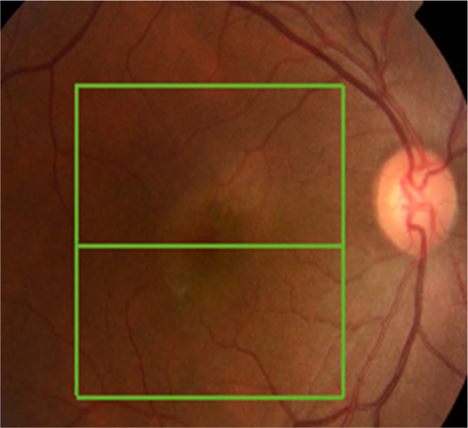
Slit lamp examination and colour fundus photography – are helpful in recognising serous retinal detachments and RPE mottling/atrophy. Subtle fluid can be challenging to notice.

Image 1. Colour fundus photo of acute CSR. Notice RPE mottling superior to the fovea.

Optical coherence tomography, invasive angiographic tests (fluorescein angiography and indocyanine-green angiography), and golden diagnostic imaging in diagnosing and monitoring the CSR and other conditions within the spectrum.

Image 2. OCT scan of acute CSR. Presence of subretinal fluid with no drusen.
Image 3. Choroidal excavation. Note no signs of fluid and pachychoroid (increased choroidal thickness).

Fundus autofluorescence is a valuable tool for recognising the localised thickening area. Very often, extrafoveal regions that are not well seen clinically can be observed.

Image 4. Fundus autofluorescence image. It is a helpful tool for checking the areas of damaged RPE.
05

Management and treatment

Active CSR typically resolves independently, so patients are only monitored.

There is no approved treatment for CSR or other pachychoroid conditions. The laser treatment modalities are options to treat central serous retinopathy (micropulse versus half-dose PDT).(22,23)

There is no -substantial evidence of the benefits of anti-adrenergic agents in treating CSR.(24)

Anti-VEGF injections are used to treat secondary CNV.(25)

06

References

1 Lam D. et al. Central Serous Retinopathy In: Schachat AP, Wilkinson CP, Hinton DR, Sadda SR, Wiedemann P, eds. Ryan’s Retina. 6th ed.Philadelphia. Elsevier/Saunders; 2018:chap75.

2 Wang M, Munch IC, Hasler PW, Prünte C, Larsen M. Central serous chorioretinopathy. Acta ophthalmologica. 2008 Mar;86(2):126-45.

3 Guyer DR, Yannuzzi LA, Slakter JS, Sorenson JA, Hope-Ross M, Orlock DR. Digital indocyanine-green videoangiography of occult choroidal neovascularisation. Ophthalmology. 1994 Oct 1;101(10):1727-37.

4 Baran NV, Gürlü VP, Esgin H. Long‐term macular function in eyes with central serous chorioretinopathy. Clinical & experimental ophthalmology. 2005 Aug;33(4):369-72.

5 Spaide RF, Campeas L, Haas A, Yannuzzi LA, Fisher YL, Guyer DR, Slakter JS, Sorenson JA, Orlock DA. Central serous chorioretinopathy in younger and older adults. Ophthalmology. 1996 Dec 1;103(12):2070-80.

6 Kitzmann AS, Pulido JS, Diehl NN, Hodge DO, Burke JP. The incidence of central serous chorioretinopathy in Olmsted County, Minnesota, 1980–2002. Ophthalmology. 2008 Jan 1;115(1):169-73.

7 Leibowitz HM, Krueger DE, Maunder LR, Milton RC, Kini MM, Kahn HA, Nickerson RJ, Pool J, Colton TL, Ganley JP, Loewenstein JI. The Framingham Eye Study monograph: An ophthalmological and epidemiological study of cataract, glaucoma, diabetic retinopathy, macular degeneration, and visual acuity in a general population of 2631 adults, 1973-1975. Survey of ophthalmology. 1980 May 1;24(Suppl):335-610.

8 Garg SP, Dada T, Talwar D, Biswas NR. Endogenous cortisol profile in patients with central serous chorioretinopathy. British Journal of Ophthalmology. 1997 Nov 1;81(11):962-4.

9 Carvalho-Recchia CA, Yannuzzi LA, Negrão S, Spaide RF, Freund KB, Rodriguez-Coleman H, Lenharo M, Iida T. Corticosteroids and central serous chorioretinopathy. Ophthalmology. 2002 Oct 1;109(10):1834-7.

10 Tewari HK, Gadia R, Kumar D, Venkatesh P, Garg SP. Sympathetic–parasympathetic activity and reactivity in central serous chorioretinopathy: a case–control study. Investigative Ophthalmology & Visual Science. 2006 Aug 1;47(8):3474-8.

11 Prünte C, Flammer J. Choroidal capillary and venous congestion in central serous chorioretinopathy. American Journal of Ophthalmology. 1996 Jan 1;121(1):26-34.

12 Casella AM, Berbel RF, Bressanim GL, Malaguido MR, Cardillo JA. Helicobacter pylori as a potential target for the treatment of central serous chorioretinopathy. Clinics. 2012;67:1047-52.

13 Fraunfelder FW, Fraunfelder FT. Central serous chorioretinopathy associated with sildenafil. Retina. 2008 Apr 1;28(4):606-9.

14 Hassan L, Carvalho C, Yannuzzi LA, Iida T, NegrÃo S. Central serous chorioretinopathy in a patient using methylenedioxymethamphetamine (MDMA) or “ecstasy.” Retina. 2001;21(5):559–561.

15 https://eyewiki.aao.org/Central_Serous_Chorioretinopathy#cite_note-:9-24

16 Yavaş GF, Küsbeci T, Kaşikci M, Günay E, Doğan M, Ünlü M, Inan ÜÜ. Obstructive sleep apnea in patients with central serous chorioretinopathy. Current Eye Research. 2014 Jan 1;39(1):88-92.

17 Cheung CM, Lee WK, Koizumi H, Dansingani K, Lai TY, Freund KB. Pachychoroid disease. Eye. 2019 Jan;33(1):14-33.

18 Maruko I, Iida T, Ojima A, Sekiryu T. Subretinal dot-like precipitates and yellow material in central serous chorioretinopathy. Retina. 2011 Apr 1;31(4):759-65.

19 Dansingani KK, Naysan J, Bala C, Freund KB. En Face imaging of pachychoroid spectrum disorders with swept-source OCT. Investigative Ophthalmology & Visual Science. 2015 Jun 11;56(7):2787-.

20 Pang CE, Freund KB. Pachychoroid pigment epitheliopathy may masquerade as acute retinal pigment epitheliitis. Investigative ophthalmology & visual science. 2014 Aug 1;55(8):5252-.

21 Phasukkijwatana N, Freund KB, Dolz-Marco R, Al-Sheikh M, Keane PA, Egan CA, Randhawa S, Stewart JM, Liu Q, Hunyor AP, Kreiger A. Peripapillary pachychoroid syndrome. Retina. 2018 Sep 1;38(9):1652-67.

22 Nicoló M, Eandi CM, Alovisi C, Grignolo FM, Traverso CE, Musetti D, Piccolino FC. Half-fluence versus half-dose photodynamic therapy in chronic central serous chorioretinopathy. American Journal of Ophthalmology. 2014 May 1;157(5):1033-7.

23 Scholz P, Altay L, Fauser S. Comparison of subthreshold micropulse laser (577 nm) treatment and half-dose photodynamic therapy in patients with chronic central serous chorioretinopathy. Eye. 2016 Oct;30(10):1371-7.

24 Lotery A, Sivaprasad S, O’Connell A, Harris RA, Culliford L, Cree A, Madhusudhan S, Griffiths H, Ellis L, Chakravarthy U, Peto T. Eplerenone versus placebo for chronic central serous chorioretinopathy: the VICI RCT.

25 Scholz P, Altay L, Fauser S. Comparison of subthreshold micropulse laser (577 nm) treatment and half-dose photodynamic therapy in patients with chronic central serous chorioretinopathy. Eye. 2016 Oct;30(10):1371-7.