Choroidal nevus

Referral priority: Low or urgent – depending on clinical signs.

Choroidal nevi might require regular follow-up to detect a malignant transformation. Follow-up or refer according to local guidelines. Clinical signs determine the priority for a referral.

Click on one of the cards below to learn about the specific eye condition.

Written by
Marko Lukic
Edited by
Svein Tindlund and Jon Gjelle
Published
June 2023

Sections
01
Introduction

Read more
02
Symptoms & clinical signs

Read more
03
Diagnostic procedures

Read more
04
Management and treatment

Read more
05
References

Read more
01

Introduction

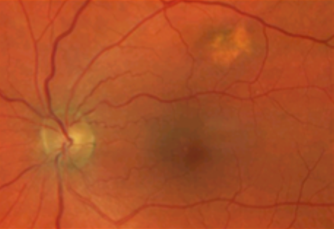
Choroidal nevi are common tumours/choroidal lesions composed of collections of benign-appearing uveal melanocytes called nevus cells.(1,2) In most cases, nevi represent benign lesions that require no treatment or additional workup. Due to the risk of transforming into a choroidal melanoma, those lesions require regular monitoring and being familiar with suspected features is crucial.

Greenstein et al. found an overall prevalence of 2.1% for choroidal nevi in a mixed population, with the highest prevalence in Caucasians (4.1%).(3) Histological findings have demonstrated that the prevalence of choroidal nevi may be higher than what is observed clinically.(4)

Thirty-one per cent of nevi will grow over time (years to decades) without malignant transformation. -According to Shields et al., the transformation of choroidal nevi is generally rare, occurring at a rate of 1 in 8,845 nevi cases per year.(5) Fair skin, the ability to tan, and light eye colour are statistically important risk factors – of choroidal melanoma transformation (6)

02

Symptoms & clinical signs

Choroidal nevi are clinically presented as flat or slightly elevated pigmented lesions with defined but not well-demarcated margins. They are often located within the posterior pole and usually do not cause any visual disturbances. The size of the -nevus is compared – to the diameter of the optic disc (approximately 1.5 mm), and it may be from one-third to seven disc diameters (DD). In sporadic cases, the -nevus can be larger than 10 mm in basal diameter, which, according to Blue Mountain Eye Study, is only the case in 1.5% of choroidal nevi.(7,8) Such nevi are called giant choroidal nevi. Then, the choroidal nevi are no thicker than 2 mm, and the pigmentation of the lesion may vary. Some nevi may be heavily pigmented, while others may be amelanotic. According to the report from Shields et al., around 10% of patients have a melanocytic-nevus.(9) Some nevi may represent an amelanotic halo surrounding the lesion.

The presence of drusen and a halo of depigmentation around a pigmented choroidal lesion (nevus) are reassuring findings most widely observed in choroidal nevi rather than melanomas. In rare cases, there may be atrophy or hyperplasia of the RPE, including subretinal fluid. As you can see below, subretinal fluid is always a suspected sign, and ophthalmologists and ocular oncologists must see those patients. However, in some cases, the chronic damage of RPE in non-malignant choroidal lesions may lead to subretinal fluid. If the fluid affects the fovea, then patients become symptomatic. In even fewer cases, the choroidal nevus may develop a secondary choroidal neovascular membrane. If the membrane affects the vision, it should be treated with intravitreal anti-VEGF injections.(10)

How to recognise choroidal melanoma from choroidal nevi

Shields and colleagues analysed 2,514 cases of choroidal nevi to determine predictive features of growth into melanoma. Using the data obtained in this study, they developed a mnemonic for these predictive features: “To Find Small Ocular Melanoma Using Helpful Hints Daily.”11

  • T: Thickness > 2 mm
  • F: Fluid (subretinal)
  • S: Symptoms (decreased vision, flashes, or floaters)
  • O: Orange pigment (presence of lipofuscin)
  • M: Margin within 3 mm of the optic disc (2 discs diameters from the optic disc)
  • UH: Ultrasonographic hollowness
  • H: Absence of surrounding halo. Nevi usually show a surrounding clear halo consisting of an atrophied retina.
  • D: Absence of drusen.


Image 1. The image represents a suspected choroidal nevus. Please, note that nevus is elevated and has drusen on its surface—the suspected presence of subretinal fluid.
Image 2. The OCT scan done over the nevus revealed the presence of subretinal fluid and drusen. There are no signs of lipofuscin or shaggy photoreceptors. According to the criteria, it comes to only one risk factor. The lesion represents a nevus and requires monitoring by an ophthalmologist.

Choroidal melanocytic tumours that display no risk factors have a 3% chance -of growth at the five-year mark and most likely represent choroidal nevi. Tumours that express one risk factor have a 38% chance of growth, and those with two or more risk factors, growth is observed in over 50% of cases at the five- year mark.

03

Diagnostic procedures

Fundus photography – is used to identify chorioretinal lesions and monitor them. Be aware that scanning laser ultra-widefield cameras represent pseudo-coloured photos, and the actual colour of the lesion on other machines or clinically on a slit lamp may look different.

Image 3. A flat, juxta papillary choroidal nevus.
Image 4. Amelancotic choroidal nevus.

Ultrasound and colour Doppler are important and, in most cases, primary diagnostic tools to diagnose choroidal melanoma. The most common representation of the tumour is a dome-shaped choroidal elevation. Other features that may be present are an acoustically hollow zone within the tumour, choroidal excavation, and subretinal fluid. The colour doppler confirms internal blood flow, which is not true with nevi.


  Apical height  Largest basal diameter 
Small  1.0 – 2.5 mm  5.0 – 16.0 mm 
Medium  2.5 – 10 mm  less than 16 mm 
Large  more than 10 mm  more than 16 mm 

Table 1. The table displays tumour size-modified criteria as per Collaborative Ocular Melanoma Study

Spectral-domain OCT – is used to identify whether the pigmented lesion is elevated or flat and to reveal the presence of subretinal fluid, drusen, and lipofuscin.

Image 5. The OCG image of a flat choroidal naevus with overlying drusen. No signs of subretinal fluid or lipofuscin.

Enhanced Depth Imaging Spectral-Domain OCT (EDI OCT) – this imaging modality evaluated deeper structures like choroids better than the standard OCT imaging. It may better visualise shaggy photoreceptors, lipofuscin deposits, and thinning of choriocapillaris.(10)

Fundus autofluorescence – is used to identify lipofuscin. Lipofuscin is present in suspicious nevi and many melanomas. Lipofuscin fluorescence is brighter than drusen (drusen are typical in choroidal nevi).

04

Management and treatment

Choroidal nevi that do not raise any suspicion do not need any treatment. However, it is typically advised to keep a yearly check on them. For instance, schedule another one during your first meeting with the patient after six months. If the lesion remains unchanged, observe the patient every year.

If the patient has suspected features, refer the patient urgently to a local ophthalmologist. Such patients will be monitored on a 6-monthly basis and have multi-modal imaging to observe or discover potentially sinister features.

05

References

1 Green W. Uveal tract. Spencer W. Ophthalmic pathology: an atlas and text. WB Saunders: Philadelphia; 1997.

2 Zimmerman LE. Melanocytes, melanocytic nevi, and melanocytomas: the Jonas S. Friedenwald Memorial Lecture. Investigative Ophthalmology & Visual Science. 1965 Feb 1;4(1):11-41.

3 Greenstein MB, Myers CE, Meuer SM, Klein BE, Cotch MF, Wong TY, Klein R. Prevalence and characteristics of choroidal nevi: the multi-ethnic study of atherosclerosis. Ophthalmology. 2011 Dec 1;118(12):2468-73.

4 Naumann G. Pigmentierte naevi der Aderhaut und des Ciliarkörpers. Adv Ophthalmol. 1970;23:187-272.

5 Shields CL, Furuta M, Berman EL, Zahler JD, Hoberman DM, Dinh DH, Mashayekhi A, Shields JA. Choroidal nevus transformation into melanoma: analysis of 2514 consecutive cases. Archives of Ophthalmology. 2009 Aug 1;127(8):981-7.

6 Weis E, Shah CP, Lajous M, Shields JA, Shields CL. The association between host susceptibility factors and uveal melanoma: a meta-analysis. Archives of ophthalmology. 2006 Jan 1;124(1):54-60.

7 Li HK, Shields CL, Mashayekhi A, Randolph JD, Bailey T, Burnbaum J, Shields JA. Giant choroidal nevus: clinical features and natural course in 322 cases. Ophthalmology. 2010 Feb 1;117(2):324-33.

8 Sumich P, Mitchell P, Wang JJ. Choroidal nevi in a white population: the Blue Mountains Eye Study. Archives of Ophthalmology. 1998 May 1;116(5):645-50.

9 Shields CL, Furuta M, Mashayekhi A, Berman EL, Zahler JD, Hoberman DM, Dinh DH, Shields JA. Clinical spectrum of choroidal nevi based on age at presentation in 3422 consecutive eyes.
Ophthalmology. 2008 Mar 1;115(3):546-52.

10 Mines JA, Freilich DB, Friedman AH, Lazar M. Choroidal (subretinal) neovascularization secondary to choroidal nevus and successful treatment with argon laser photocoagulation. Ophthalmologica. 1985;190(4):210-8.

11 Shields CL, Furuta M, Berman EL, Zahler JD, Hoberman DM, Dinh DH, Mashayekhi A, Shields JA. Choroidal nevus transformation into melanoma: analysis of 2514 consecutive cases. Archives of Ophthalmology. 2009 Aug 1;127(8):981-7.