Contact lens complications

Referral priority: Routine or urgent

Urgently refer all patients with signs of microbial keratitis to an ophthalmologist or a hospital, following local guidelines.

Written by
Marko Lukic
Edited by
Svein Tindlund and Jon Gjelle
Published
June 2023

Sections
01
Introduction

Read more
02
Symptoms

Read more
03
Clinical signs

Read more
04
Diagnostic procedures

Read more
05
Management and treatment

Read more
06
References

Read more
01

Introduction

Contact lens wearing is safe; about 140 million people worldwide wear them.(1,2) It is important to remember that several serious complications – occur if proper care is neglected – The complications are directly induced or aggravated by contact lens wear. The mechanisms by which contact lenses induce alterations are: trauma, decreased corneal oxygenation, reduced corneal and conjunctival lubrication, stimulation of allergic and inflammatory responses, and infection.3

Common complications may arise, such as discomfort, dry eye, corneal discomfort, or giant cell conjunctivitis. Furthermore, it is important to be aware of serious sight-threatening complications such as – corneal neovascularisation, corneal abrasion, or infections keratitis (with or without a corneal ulcer).(4) In addition, over-extending of contact lens wearing may induce contact lens-induced acute red eye (CLARE) syndrome.(5)

Microbial keratitis is one of the most severe complications of contact lens wear. Keratitis results from an alteration in the cornea’s defence mechanisms, allowing bacteria to invade when an epithelial defect is present. Overnight use is a leading risk factor as is extended-wear poor hygiene and not using appropriate cleaning solutions.(6,7) Organisms responsible include pseudomonas aeruginosa, staphylococcus, streptococcus, serratia, and acanthamoeba. The risk for acanthamoeba infection is water and infected lens solutions.

Wearing contacts reduces the amount of oxygen that the cornea receives from the surface of the eye (hypoxia), which can lead to cornea swelling (corneal oedema). Over time, the cornea tries to get more oxygen by growing new blood vessels (corneal neovascularisation). If severe, the vessels can grow into the centre of the cornea and cause vision loss.

Sterile corneal infiltrates usually appear on the peripheral cornea and are secondary to contact lens wear and/or bacterial endotoxins. They may represent a diagnostic dilemma for keratitis.

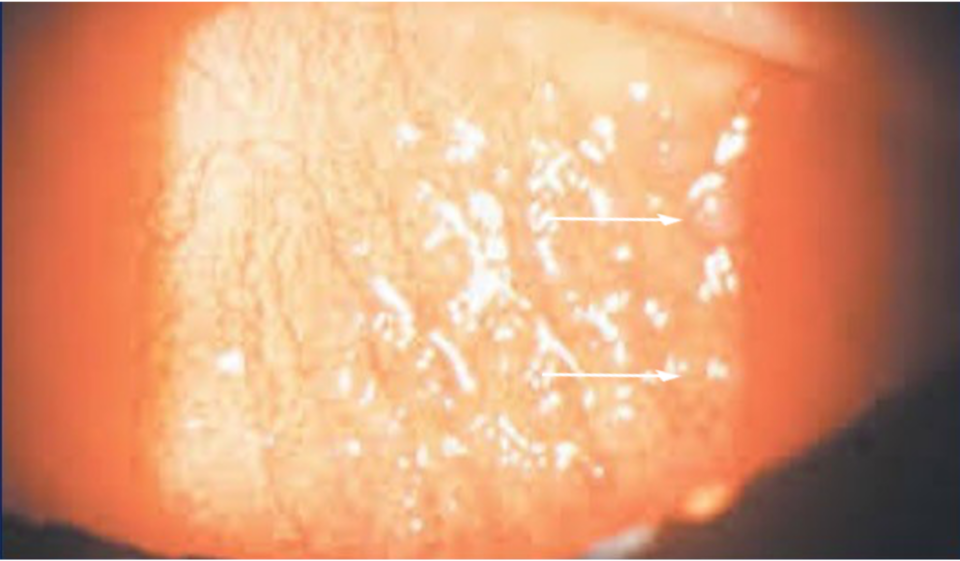
Giant-cell conjunctivitis (GPC) can develop in the upper eyelid due to continuous rubbing against the contact lens. If not detected and treated promptly, patients may experience contact lens intolerance.

Maintaining proper contact lens hygiene is crucial. This involves refraining from wearing contacts while sleeping, showering, or swimming to minimise the chances of experiencing severe complications. In addition, opting for daily disposable contact lenses can significantly reduce the risk of developing infectious keratitis.(4)

02

Symptoms

Various symptoms may appear in complications related to contact lenses. It is important to be familiar with warning symptoms like redness or irritation, eye pain, sensitivity to light, blurry or worsening vision, excessive tearing, or discharge. Those signs may indicate infectious keratitis

Acanthamoeba keratitis is identified by the presence of disproportionately severe pain compared to the observed clinical findings. In one study, 95% of patients complained of pain. Patients may also complain of decreased vision, redness, foreign body sensation, photophobia, tearing, and discharge. Symptoms may wax and wane; they may be pretty severe at times.(8)

Patients with lens-induced conjunctivitis may feel foreign body sensations and itchiness.

Inquire about the patient’s contact lens usage, including the type of lenses they use, the duration since their last lens replacement, the number of hours they wear the lenses continuously, and their cleaning routine. Also, ask whether they swim or shower while wearing contact lenses.

03

Clinical signs

Contact lens wearing may affect corneal integrity, which may cause corneal defects such as punctate keratopathy, corneal abrasions, foreign body tracks, corneal dellen (shallow, saucer-like excavations at the margin of the cornea)(9), and microcysts.

Corneal infiltrate is usually present with microbial keratitis. However, remember that corneal ulcer (open sore of the cornea) is not necessarily present with microbial keratitis.

Early signs of Acanthamoeba keratitis may be mild and non-specific. Possible findings include epithelial irregularities, epithelial or anterior stromal infiltrates, and pseudodendrites. Deep stromal infiltrates (ring-shaped, nummular, or disciform), satellite lesions, and persistent corneal defects are grouped among the late signs of the condition.

Redness of the eye may indicate CLARE syndrome or giant cell conjunctivitis. Patients with conjunctivitis may also have swollen and droopy eyelids and enlarged papillae.

Image 1. A contact lens-induced acute red eye (CLARE) syndrome in a lens wearer.
Image 2. An eye with acute microbial keratitis in a contact lens wearer. Inflamed conjunctiva with localised corneal staining and white stromal infiltrate.
Image 3. Enlarged papillae of the superior eyelid in a contact lens wearer.
04

Diagnostic procedures

A thorough eye examination is vital in making a proper diagnosis. It is crucial to be familiar with symptoms and clinical signs.

Corneal culture is a diagnostic procedure for identifying an infectious agent. If the ulcer is large, central, unresponsive to current treatment, or there is suspicion of an atypical infectious organism, it may be necessary to consider culturing.(10,11) The corneal swab needs to be done by an experienced physician. Avoid corneal ulcer base if there is significant corneal thinning. It is also best to avoid obtaining only purulent material, as it is unlikely to yield a positive result.(10,12)

05

Management and treatment

Patients with contact lens complications should be advised to stop wearing contact lenses until symptoms improve.

If available, consider prescribing the patient preventive antibiotic therapy and lubricant eye drops in case of corneal defects like corneal abrasions. Consider re-fitting of the contact lens after healing.

Advise patients with corneal oedema secondary to lens overwear to reduce lens wearing and find higher oxygen permeable lenses.

Closely monitor patients with sterile corneal infiltrates. It is recommended to prescribe prophylactic antibiotic therapy followed by local steroids.

Combination mast cell stabilisers/antihistamines and modification of contact lens type/hygiene are the primary treatments for GPC. Topical steroids can be used to treat severe GPC but are not always necessary, especially in mild cases.

Corneal ulcers that are not sight-threatening are empirically treated with fluoroquinolone agents. Vision-threatening corneal cultures and Gram stain should be performed, and broad-spectrum antibiotics and cycloplegics should be initiated while culture and sensitivity results are pending. Traditionally, Vancomycin and fortified aminoglycosides are prescribed hourly.

Treatment of acanthamoeba keratitis involves a multi-drug regimen of anti-amebic drugs, which include polyhexamethylene biguanide, propamidine isethionate, and neomycin.

06

References

1https://www.cdc.gov/contactlenses/index.html#:~:text=While%20contact%20lenses%20are%20usually,infections%2C%20such%20as%20microbial%20keratitis.

2 Cope JR, Collier SA, Nethercut H, Jones JM, Yates K, Yoder JS. Risk behaviors for contact lens–related eye infections among adults and adolescents—United States, 2016. Morbidity and Mortality Weekly Report. 2017 Aug 8;66(32):841.

3 https://eyewiki.aao.org/Contact_Lens_Complications

4 https://jamanetwork.com/journals/jama/fullarticle/2779826

5 Fagan XJ, Jhanji V, Constantinou M, Amirul Islam FM, Taylor HR, Vajpayee RB. First contact diagnosis and management of contact lens-related complications. International ophthalmology. 2012 Aug;32(4):321-7.

6 Cheung N, Nagra P, Hammersmith K. Emerging trends in contact lens-related infections. Current opinion in ophthalmology. 2016 Jul 1;27(4):327-32.

7 Stapleton F, Carnt N. Contact lens-related microbial keratitis: how have epidemiology and genetics helped us with pathogenesis and prophylaxis. Eye. 2012 Feb;26(2):185-93.

8 https://eyewiki.aao.org/Acanthamoeba_Keratitis

9 Fuchs A. Pathological dimples (“Dellen”) of the cornea. American Journal of Ophthalmology. 1929 Nov 1;12(11):877-83.

10 Matoba AY, Harris DJ, Mark DB, Meisler DM. American Academy of Ophthalmology Cornea/External Disease Panel, Preferred Practice Patterns Committee. Blepharitis. American Academy of Ophthalmology, San Francisco, CA. 2003.

11 Gerstenblith A, Rabinowitz M. Bacterial keratitis and corneal culture procedure. The Wills Eye Manual: Office and Emergency Room Diagnosis and Treatment of Eye Disease. 6th ed. Philadelphia: Wolters Kluwer/Lippincott Williams & Wilkins;2012:69-73, 435-437.

12 Kanski J, Bowling B. Bacterial Keratitis. Clinical Ophthalmology: A Systematic Approach. 7th ed. Edinburgh: Butterworth-Heinemann/Elsevier, 2011:173-80.