Diabetic retinopathy

Referral priority: From mild to urgent

Urgently refer all patients with signs of proliferative disease to an ophthalmologist or the hospital following local guidelines. Semi-urgently refer patients with all non-proliferative forms and the presence of diabetic macular oedema, as well as those with severe non-proliferative diabetic retinopathy, to an ophthalmologist according to local guidelines.

Written by
Marko Lukic
Edited by
Svein Tindlund and Jon Gjelle
Published
June 2023

Sections
01
Introduction

Read more
02
Symptoms

Read more
03
Clinical signs

Read more
04
Diagnostic procedures

Read more
05
Management and treatment

Read more
06
References

Read more
01

Introduction

The pandemic of diabetes mellitus represents a substantial global health challenge. Diabetes causes many long-term systemic complications that considerably impact the patient and society, as it typically affects individuals in their most productive years.(1) There are two types of diabetes, type 1 and 2. Type 1 diabetes mellitus (T1DM) is characterised by the destruction of beta cells in the pancreas by an autoimmune mechanism, whereas type 2 diabetes mellitus (T2DM) is a relationship between lifestyle and genetics.

The International Diabetes Federation published data that indicate that the prevalence of diabetes mellitus in Europe is 8.5% of people aged between 20 and 79 years, which means that 33 million people in the European Union are affected by this silent and severe condition.(2) The total number of people diagnosed with diabetes mellitus worldwide is projected to rise to 366 million in 2030.(3)

Diabetic retinopathy represents microvascular end-organ damage as a result of diabetes. The Wisconsin Epidemiologic Study of Diabetic Retinopathy (WESDR) Cohort showed that after 20 years of diabetes mellitus, 99% of patients with type 1 and 60% of patients with type 2 show some retinopathy.(4) Furthermore, a meta-analysis of 14 prospective studies showed that 1 in 4 with diabetes develop diabetic retinopathy within the mean follow-up period of 5.7 years, whilst another meta-analysis of 35 prospective studies revealed an incidence of DR among people with diabetes of 34.6%.(5,6)

There are established risk factors for developing diabetic retinopathy; diabetes duration, uncontrolled blood pressure and blood sugar levels, hypertension, dyslipidaemia, ethnicity, pregnancy, and smoking.(7) The role of glycated haemoglobin (HbA1c) in its correlation with the risk of developing and progressing diabetic retinopathy has been well established. The HbA1c is an important indicator of long-term glycemic control to reflect the cumulative glycemic history of the preceding two to three months.(8) There are two scales of HbA1c used in everyday clinical practice. The targets of diabetic patients under the old classification were between 6.5 and 7.5%, while the new measurement indicates 48-59 mmol/mol.(9) As per Diabetes Control and Complications Trial (DCCT), and later on confirmed by The Epidemiology of Diabetes Interventions and Complications Study (EDICS), the intensive control of blood glucose levels, where the mean HbA1c was 7.2%, achieved a decrease in the incidence of diabetic retinopathy by 76% and slowing the progression of diabetic retinopathy by 54%.(10,11)

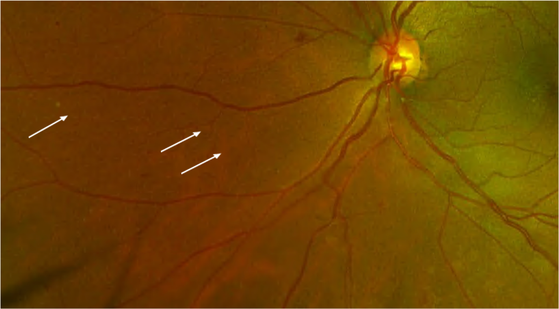
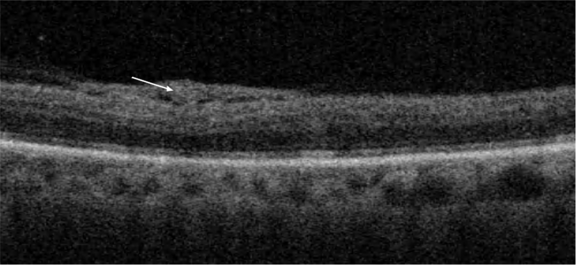
Diabetic retinopathy ranges from non-proliferative diabetic retinopathy (NPDR) and its stages to proliferative diabetic retinopathy (PDR). The proliferative stage is defined by the presence of new vessels of the retina, optic disc, and/or iris and iridocorneal angle. As the disease progresses, associated diabetic macular oedema may also become apparent. Three major events are involved in the pathomechanism of diabetic retinopathy and, consequently, in diabetic macular oedema; capillary leakage, capillary occlusion, and retinal ischaemia with its sequelae.(7) On the molecular level, hyperglycemia leads to the activation of alternative pathways of glucose metabolism, such as the polyol pathway , advanced glycation endproducts (AGEs) formation, protein kinase C (PKC) activation, hexosamine pathway flux, and Poly(ADP-ribose) polymerase activation.(12,13) The result of these pathways is the activation of cytokines and growth factors, leading to vascular endothelial dysfunction, increased vascular permeability, and eventual microvascular occlusion.(12) Microvascular occlusion leads to retinal ischemia, which promotes neovascularisation and the formation of IRMAs.(12)

02

Symptoms

Patients with early stages of diabetic retinopathy and without macular oedema may be asymptomatic. Those with more advanced settings may experience a progressive decrease in vision (due to macular oedema and/or macular ischaemia), floaters (secondary to vitreous haemorrhage), and visual fields defect (in the event of tractional retinal detachment).(7)

Remember that patients may have fluctuating vision due to changes in blood sugar levels and the degree of macular oedema. Both high and low blood sugar levels may cause changes in the crystalline lens, which causes a difference in the quality of vision.

Always take a thorough medical history, including blood sugar and blood pressure control questions. Remind your patients of the importance of strict control of the metabolic status and being familiar with HbA1c.

03

Clinical signs

It is vital to be familiar with the clinical features of diabetic retinopathy and the grades of the disease. The clinical signs that may be found in diabetic retinopathy are as follows:

Non-proliferative features
Microaneurysms are the earliest clinical sign of diabetic retinopathy and occur secondary to capillary wall outpouching due to pericyte loss. They appear as small red dots in the superficial retinal layers.

Retinal haemorrhages may appear as a dot, blot, or flame-shaped haemorrhage. – Location and appearance determine them. Dot and blot haemorrhages occur as microaneurysms rupture in the deeper layers of the retina, such as the inner nuclear and outer plexiform layers. Dot haemorrhages may be like microaneurysms. Flame-shaped haemorrhages are splinter haemorrhages in the more superficial nerve fibre layer.

Retinal/macular oedema and hard exudates are caused by the breakdown of the blood-retina barrier, allowing leakage of serum proteins, lipids, and protein from the vessels.

The occlusion of precapillary arterioles causes cotton-wool spots, which are nerve fibre layer infarctions.

Venous beading is when the walls of prominent retinal veins lose their normal parallel alignment and appear more like a string of sausages. They reflect increased/progression of retinal ischaemia.

Image 1. Venous beading in a patient with severe non-proliferative diabetic retinopathy. Note the parallel alignment and appearance of a string of sausages.

Venous loops and venous reduplication represent shunts that bypass non-thrombotic occlusion of major retinal veins.(14)

Intraretinal microvascular abnormalities (IRMAs) are remodelled capillary beds without proliferative changes. They are – typically located on the borders between perfused and non-perfused retinas.

Image 2. Intraretinal microvascular abnormality (IRMA) indicates an area of a significant drop in retinal perfusion.

Proliferative features
New vessels are the hallmark of proliferative diabetic retinopathy. They may be on the surface or near the optic disc and then are called new vessels of the disc (NVD). Or they may be present within 3 disc diameters (DD) of major retinal vessels and are – referred to as new vessels elsewhere (NVE). More advanced cases may present with new vessels of the iridocorneal angle and/or new vessels of the iris.

Image 3. New vessels of the retina (NVEs) in the inferonasal quadran.

Preretinal haemorrhage is one of the features of advanced proliferative diabetic retinopathy and represents a haemorrhage in the space between the hyaloid and internal limiting membranes.

Image 4. Inferior preretinal and vitreous haemorrhage. Note tiny but abnormal vessels on the surface of the optic disc (NVDs).

Vitreous haemorrhage means the presence of blood in the vitreous gel.

Fibrovascular complexes appear on the retinal surface and may be more vascular or fibrotic, depending on the perfusion of the vessels within the complex. They may cause tractional retinal detachment.

Grading stages of diabetic retinopathy:

  • Non-proliferative diabetic retinopathy
  • Mild– the presence of a few microaneurysms
  • Moderate – the presence of microaneurysms, dot and blot haemorrhages. More advanced stages may have the presence of venous beading in one quadrant.
  • Severe follows the so-called “4-2-1” rule: four quadrants of diffuse retinal haemorrhages and microaneurysms, two or more quadrants of venous beading, or one or more quadrants of Intraretinal Microvascular Abnormalities (IRMA).

Proliferative diabetic retinopathy:

Early or high-risk proliferative diabetic retinopathy (PDR) classifies it.(15)
NVD is one-third to one-half, or greater, of the disc area (DA)
There may be any amount of NVD with vitreous or preretinal haemorrhage
NVE is one-half or greater of the DA, with preretinal or vitreous haemorrhage

04

Diagnostic procedures

Slit lamp examination and colour fundus photography are classical examination methods where it is important to be familiar with the clinical signs mentioned above.

Optical coherence tomography – it is a handy tool to srecognise and monitor diabetic macular oedema, recognise signs of diabetic maculopathy, and distinguish IRMAs from new vessels (active new vessels are located above the internal limiting membrane when IRMAs are within retinal tissue).

Image 5. OCT scans over a complex of new vessels. Note hyperreflective material above the ILM, adjacent to the posterior hyaloid membrane.

OCT angiography is a modern tool that is used to assess macular and retinal perfusion. Be aware that it is not the same tool as fluorescein angiography, and important essential differences exist between those two modalities. Nonetheless, it is a non-invasive tool and helps to quickly investigate retinal perfusion and/or the presence of new vessels.

05

Management and treatment

Patients must keep their metabolic status under control as best as possible. The role of eye care specialists is to advise patients on the importance of control and potential consequences in case of progressive disease.

Two major complications of diabetic retinopathy may occur, which are essential to recognise and treat in time: diabetic macular oedema and proliferative diabetic retinopathy.

Treatment of diabetic macular oedema is nowadays based on intravitreal treatment (anti-VEGF agents and steroids). However, focal or modified macular grid lasers still have an important role.

Pan-retinal photocoagulation (PRP) is the immediate treatment for patients with proliferative diabetic retinopathy (PDR).. The laser is used to obliterate some of the ischemic peripheral retinae to decrease VEGF release and induce regression of neovascularisation. Some clinicians use intravitreal anti-VEGF injections first to cause a reduction of new vessels. DRCR protocol S showed that the anti-VEGF drug ranibizumab was non-inferior to PRP in managing patients with PDR.(16) – Patients receiving anti-VEGF injections alone require crucial follow-up. This therapy does not seem to achieve long-term involution of neovascularisation after discontinuing the injections, whereas PRP generally yields long-lasting effects. In rubeosis and neovascular glaucoma, anti-VEGF injections may be used with PRP.

Patients with very severe NPDR may also be offered PRP laser as prophylactic treatment.

Patients with non-clearing vitreous haemorrhage and fovea-involving or fovea-threatening tractional retinal detachment require vitrectomy.

06

References

1Federman JL, Gouras P, Schubert H, Slusher MM, Vrabec TRPodos SM, Yanoff M. Systemic diseases. Retina and vitreous: Textbook of Ophthalmology. 1994;9:7-24.

2 https://idf.org/our-network/regions-members/europe/welcome.html

3 Whiting DR, Guariguata L, Weil C, and Shaw J. IDF diabetes atlas: global estimates of the prevalence of diabetes for 2011 and 2030. Diabetes research and clinical practice. 2011; 94: 311-21.

4 Klein R, Klein BE, Moss SE, Davis MD, DeMets DL. The Wisconsin Epidemiologic Study of Diabetic Retinopathy: III. Prevalence and risk of diabetic retinopathy when the age at diagnosis is 30 or more years. Archives of ophthalmology. 1984 Apr 1;102(4):527-32.

5 Sabanayagam C, Banu R, Chee ML, Lee R, Wang YX, Tan G, Jonas JB, Lamoureux EL, Cheng CY, Klein BE, Mitchell P. Incidence and progression of diabetic retinopathy: a systematic review. The lancet Diabetes & endocrinology. 2019 Feb 1;7(2):140-9.

6 Yau JW, Rogers SL, Kawasaki R, Lamoureux EL, Kowalski JW, Bek T, Chen SJ, Dekker JM, Fletcher A, Grauslund J, Haffner S. Global prevalence and major risk factors of diabetic retinopathy. Diabetes care. 2012 Mar 1;35(3):556-64.

7 https://eyewiki.aao.org/Diabetic_Retinopathy

8 Sherwani SI, Khan HA, Ekhzaimy A, Masood A, Sakharkar MK. Significance of HbA1c test in diagnosis and prognosis of diabetic patients. Biomarker insights. 2016 Jan;11:BMI-S38440.

9 https://www.iddt.org/news/new-hba1c-units

10 Nathan DM, Genuth S, Lachin J, Cleary P, Crofford O, Davis M, Rand L, Siebert C. The effect of intensive treatment of diabetes on the development and progression of long-term complications in insulin-dependent diabetes mellitus. The New England Journal of Medicine. 1993 Sep 1;329(14):977-86.

11 Nathan DM, DCCT/Edic Research Group. The diabetes control and complications trial/epidemiology of diabetes interventions and complications study at 30 years: overview. Diabetes care. 2014 Jan 1;37(1):9-16.

12 Shukla UV, Tripathy K. Diabetic Retinopathy. [Updated 2021 Feb 14]. In: StatPearls [Internet]. Treasure Island (FL): StatPearls Publishing; 2021 Jan-.

13 Whitehead M, Wickremasinghe S, Osborne A, Van Wijngaarden P, Martin KR. Diabetic retinopathy: a complex pathophysiology requiring novel therapeutic strategies. Expert opinion on biological therapy. 2018 Dec 2;18(12):1257-70.

14 Bek T. Venous loops and reduplications in diabetic retinopathy. Prevalence, distribution, and pattern of development. Acta ophthalmologica Scandinavica. 1999 Apr 1;77(2):130-4.

15 Early Treatment Diabetic Retinopathy Study Research Group. Early photocoagulation for diabetic retinopathy: ETDRS report number 9. Ophthalmology. 1991 May 1;98(5):766-85.

16 Sun JK, Glassman AR, Beaulieu WT, Stockdale CR, Bressler NM, Flaxel C, Gross JG, Shami M, Jampol LM, Diabetic Retinopathy Clinical Research Network. Rationale and application of the protocol Santi–vascular endothelial growth factor algorithm for proliferative diabetic retinopathy. Ophthalmology. 2019 Jan 1;126(1):87-95.