Dry eye disease

Referral priority: Routine or moderate

Treat all patients with dry eyes according to the severity of the condition and the cause. Refer patients who require more advanced treatment to a relevant specialist following local guidelines.

Written by
Marko Lukic
Edited by
Svein Tindlund and Jon Gjelle
Published
June 2023

Sections
01
Introduction

Read more
02
Symptoms

Read more
03
Diagnostic procedures

Read more
04
Management and treatment

Read more
05
References

Read more
01

Introduction

Dry eye disease (known as dry eye syndrome, keratoconjunctivitis sicca) is a multifactorial ocular surface disease due to a loss of tear film homeostasis.(1) It is one of the most common reasons to see an eye specialist, and its prevalence varies from 7.4%- 33.7%.2 It is more common in women.(3)

The tear film covers the ocular surface and is essential for protecting the eye from the environment, lubricating the ocular surface, maintaining a smooth surface for light refraction, and preserving the health of the conjunctiva and the avascular cornea.(4) It is also an interface between the tear film and the air and is responsible for a significant amount of the eye’s focusing power.(5) The tear film is heterogeneous and is classically divided into three layers – inner mucin, middle aqueous, and outer lipid layer. The mucin layer is formed by glycoproteins secreted mainly by the goblet cells in the conjunctival epithelium, and to a lesser extent, by the acinar cells of the lacrimal gland, epithelial cells in the cornea, and conjunctiva.(6) It functions in stabilising the aqueous layer. The aqueous layer is essential for maintaining lubrication and ocular surface protection. It contains proteins, metabolites, inorganic salts, glucose, oxygen, and electrolytes (magnesium, bicarbonate, calcium, urea) essential for maintaining the ocular surface’s health and flushing away debris and debris toxins.(7) The lipid layer at the environmental-tear interface is essential for delaying the tear evaporation rate. This superficial lipid layer contains cholesterol, wax esters, fatty acids, and phospholipids produced by meibomian glands.(7)

Any condition that may affect any tear film layers (as a primary or secondary cause) may cause dry eye disease. The International Dry Eye WorkShop II (DEWS II) classifies dry eye disease into the following two major subtypes.(8)

  • Aqueous deficient dry eye (ADDE)
  • Evaporative dry eye (EDE)

There are many causes identified in dry eye disease:

Allergies, decreased hormones associated with ageing, pregnancy and related hormonal changes, thyroid eye conditions, blepharitis, medications and supplements, Sjogren’s syndrome, Lupus, Rheumatoid arthritis, chemical injury of the eye, eye surgery, infrequent blinking,  Parkinson’s, environmental,  contact lens use, neurologic conditions,  exposure keratitis,  post-refractive surgery,  inflammatory eye conditions (uveitis), diabetes, infectious keratitis, neurotrophic keratitis, Vitamin A deficiency. (4)

The most common cause of DED is meibomian gland dysfonction (MGD). MGD affects both the quality and quantity of secreted meibum, leading to changes in tear-film composition and, as a consequence, dry eyes. (1) 
02

Symptoms

The most prominent symptom of dry eye disease is tearing. Tearing is a paradoxical reflex from the brain on the irritation to help counteract the irritation.

Other symptoms include burning, stinging, itching, gritty (sandy) filling, foreign-body sensation, discharge, frequent blinking, and crusty eyelids.

Patients may also complain of blurry vision, light sensitivity, eye redness, and pain.

Healthcare professionals can utilise symptom questionnaires to establish a diagnosis of dry eye disease, validate the need for additional interventions, evaluate treatment outcomes, and determine the severity of the disease. Some of the frequently employed and validated questionnaires include the following.(9)

  • OSDI (Ocular Surface Disease Index) is the most established study instrument.
  • The Dry Eye Questionnaire (DEQ-5) is attractive due to its short length and discriminative ability The Dry Eye Questionnaire (DEQ-5) is particularly appealing due to its concise format and ability to distinguish between different levels of dry eye symptoms effectively.
  • Consider SANDE (Symptom Assessment in Dry Eye) for repeated comfort assessment.


Ocular Surface Disease Index
The Ocular Surface Disease Index (OSDI) measures symptoms of ocular surface diseases (like dry eye syndrome), helping clinicians customise treatment based on their impact on a patient’s quality of life.(10)

  • Normal ocular surface (0-12 points)
  • Mild ocular surface disease (13-22 points)
  • Moderate ocular surface disease (23-32 points)
  • Severe ocular surface disease (33-100 points)

There are also questionnaires explicitly developed for contact lens wear, such as;

  • Contact Lens Dry Eyes Questionaire (CLDEQ)
  • 8-item Contact Lens Dry Eye Questionnaire (CLDEQ-8)
  • Contact Lens Impact on Quality of Life (CLIQ)
03

Diagnostic procedures

Corneal sensation
Corneal sensation can be measured using a cotton tip applicator or, more precisely, with a Cochet-Bonnet esthesiometer.

* Remember that severe and chronic dry eye disease may cause a reduced corneal sensation.

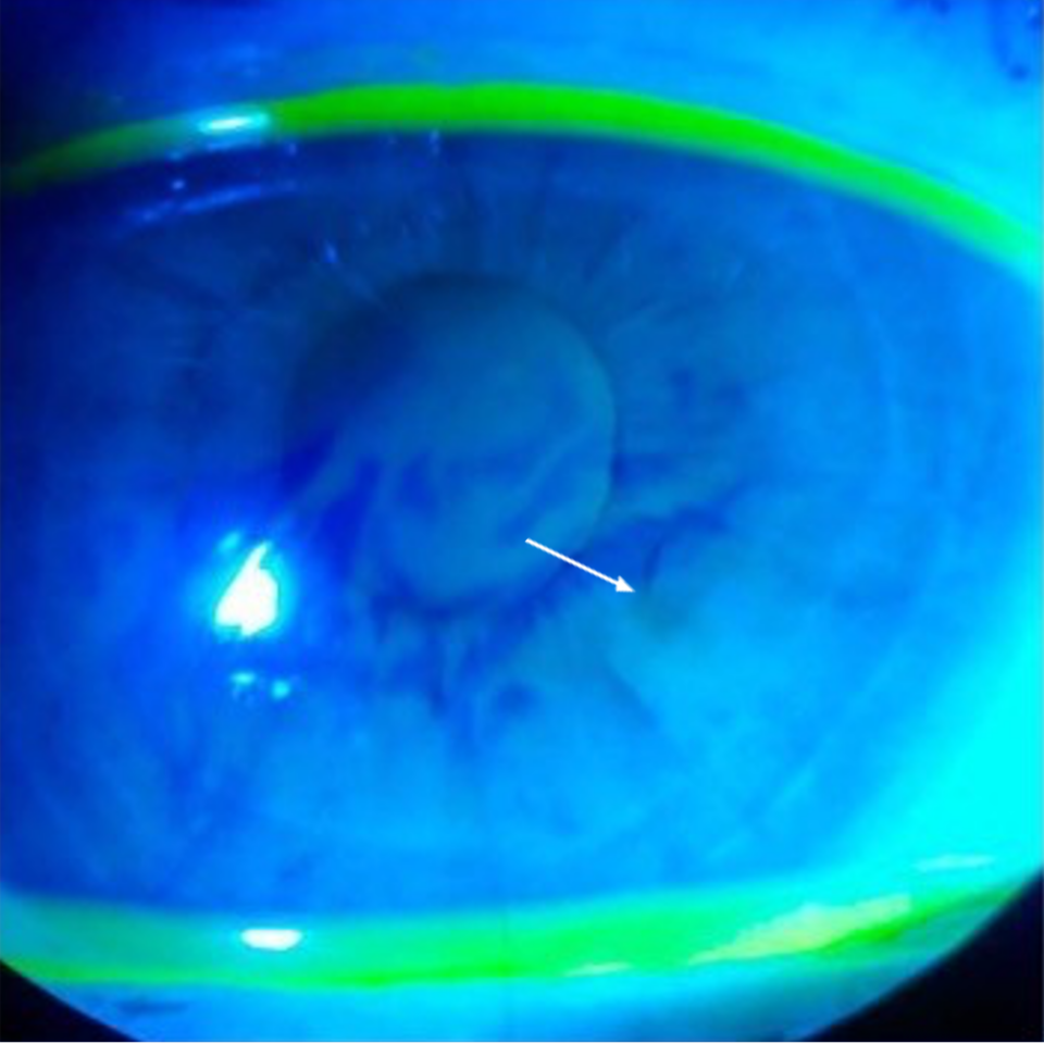
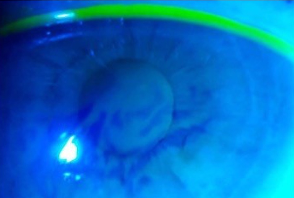
The tear break-up test (TBUT) is determined by measuring the interval between the instillation of topical fluorescein 0.5% and the appearance of the first dry spots on the cornea. Measure it before the installation of any anaesthetic eye drops. Three consecutive measurements are taken, and the median value is recorded as TBUT. A TBUT of less than 10 seconds is considered abnormal and indicative of tear instability.

Image 1. Broken tear-film (white arrow) noticeable during tear film break-up test (TBUT).

Tear film break-up time should ideally be measured non-invasively (NIBUT), e.g., using placeido ring instruments such as topographers with a dry eye module. But in lieu of this, use fluorescent tear-film break-up time (FBUT) as described above.

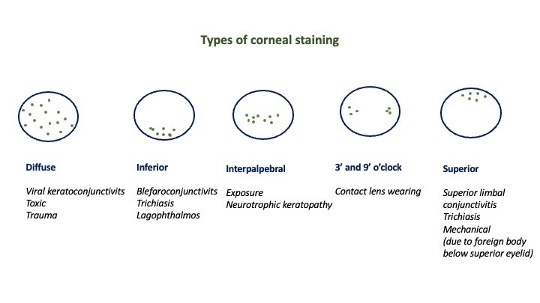
Epithelial staining
Fluorescein sodium is the most used stain in ophthalmology. Areas where the corneal or conjunctival surface epithelial cells are loose or desquamated will stain with fluorescein. Oxford Grading Scale (0-5) and National Eye Institute (NEI) scale (0-3) are the most used scales to grade fluorescein staining.

Lissamine green and Rose Bengal dyes can also be used. They are used for conjunctival staining, where lissamine green is better tolerated.

Image 2. Types of corneal staining.

Schirmer test
The Schirmer test is performed by placing a paper test strip in the lateral third of the lower eyelid after drying the inferior fornix and then measuring the length of the moistened portion of the strip after 5 minutes. The patient’s eyes are then closed for 5 minutes, and the paper strip’s wetting is measured.

The Schirmer I test is performed without anaesthesia and, thus, measures basic and reflex tearing. Less than 5 mm of wetting is abnormal; 5 to 10 mm is equivocal.

The Schirmer II test also lacks anaesthesia but is done following nasal stimulation (nasal mucosal irritation induced with a cotton tip applicator). it measures reflex tearing only and is reduced more in Sjögren’s syndrome compared to non-Sjögren’s dry eye. Wetting of less than 15 mm after 5 minutes is consistent with abnormalities of reflex secretion.

* Remember that the test is variable and has low reproducibility. It may be insensitive for mild dry eye disease.

Delayed tear clearance and tear meniscus height measurement (meniscometry)

The persistence of fluorescein dye at various time points may indicate nasolacrimal duct issues. Then, the height of the tear meniscus less than 0.25 mm is suggestive of dry eye. Medical devices automatically measure the tear meniscus, which may be useful in diagnosing dry eye syndrome.

Tear osmolarity is used to measure the osmolarity of the tear film using a device. Patients with dry eyes have increased osmolarity of their tears (hyperosmolarity). Typical values are considered to be 296±9.8 mOsm/L.(11) Greater than 308 mOsm/L indicates at least mild dry eye and has been demonstrated to be an early indicator of ocular surface instability.(12)

Tear film interferometry is a noninvasive method of grading tear film quality and estimating the thickness of the lipid layer, which is abnormal in the evaporative dry eye that is secondary to meibomian gland dysfunction.

To differentiate between MGD as a cause of DED, the quality of meibum secretion and gland expressibility should be investigated. Advanced practices may also utilise meibography to image the meibomian glands to detect potential atrophy of the glands. 

04

Management and treatment

The Management and Therapy Subcommittee of the Tear Film and Ocular Surface Society’s Dry Eye Workshop II (TFOS DEWS II) has proposed an updated (2017) algorithm to approach the treatment of dry eyes.(13)

Step 1

  • Educate the patient regarding the condition, management, and prognosis
  • Modify the patient’s local environment
  • Educate the patient on dietary modifications (including oral essential fatty acid supplementation)
  • Identify any potentially etiologic systemic/topical medications and consider modifying or eliminating offending agents.
  • Apply ocular lubricants (lipid-containing supplements in patients with meibomian gland dysfunction.
  • Institute proper lid hygiene and apply warm compresses.

Step 2 (used if the above options are inadequate)

  • Use of nonpreserved ocular lubricants to minimize preservative-induced toxicity
  • Tea tree oil therapy to treat demodicosis, if present
  • Tear conservation therapy involves procedures such as punctal occlusion and the use of moisture chamber spectacles/goggles)
  • Overnight treatments (e.g., ointment or moisture chamber devices)
  • In-office, physical heating and expression of the meibomian glands (including device-assisted therapies [e.g., LipiFlow])
  • In-office intense pulsed light therapy to treat MGD
  • Prescription drug therapy (topical antibiotic with or without steroid applied to lid margins for anterior blepharitis, limited-duration topical corticosteroid, topical secretagogues, topical nonglucocorticoid immunomodulators (e.g., cyclosporine), topical LFA-1 antagonists (e.g., lifitegrast), oral macrolide or tetracycline antibiotics

Step 3 (used if the above options are inadequate)

  • Oral secretagogue therapy
  • Application of autologous/allogeneic serum eye drops
  • Therapeutic contact lenses (soft bandage lenses, rigid scleral lenses)

Step 4 (used if the above options are inadequate)

  • Longer-duration topical corticosteroid therapy
  • Amniotic membrane grafting
  • Surgical punctal occlusion
  • Other surgical options (e.g., salivary gland transplantation, tarsorrhaphy)


05

References

1 Craig JP, Nichols KK, Akpek EK, Caffery B, Dua HS, Joo CK, Liu Z, Nelson JD, Nichols JJ, Tsubota K, Stapleton F. TFOS DEWS II definition and classification report. The ocular surface. 2017 Jul 1;15(3):276-83.

2 Alshamrani AA, Almousa AS, Almulhim AA, Alafaleq AA, Alosaimi MB, Alqahtani AM, Almulhem AM, Alshamrani MA, Alhallafi AH, Alqahtani IZ, Alshehri AA. Prevalence and risk factors of dry eye symptoms in a Saudi Arabian population. Middle East African Journal of Ophthalmology. 2017 Apr;24(2):67.

3 Rand AL, Asbell PA. Current opinion in ophthalmology nutritional supplements for dry eye syndrome. Current opinion in ophthalmology. 2011 Jul;22(4):279.

4 Chang AY, Purt B. Biochemistry, tear film. InStatPearls [Internet] 2021 Jun 15. StatPearls Publishing.

5 https://eyewiki.aao.org/Dry_Eye_Syndrome#cite_note-1

6 Hodges RR, Dartt DA. Tear film mucins: front line defenders of the ocular surface; comparison with airway and gastrointestinal tract mucins. Experimental eye research. 2013 Dec 1;117:62-78.

7 Dartt DA, Willcox MD. Complexity of the tear film: importance in homeostasis and dysfunction during disease. Experimental eye research. 2013 Dec;117:1.

8 Lemp MA, Foulks GN. The definition and classification of dry eye disease. Ocul Surf. 2007 Apr;5(2):75-92.

9 Wolffsohn JS, Arita R, Chalmers R, Djalilian A, Dogru M, Dumbleton K, Gupta PK, Karpecki P, Lazreg S, Pult H, Sullivan BD. TFOS DEWS II diagnostic methodology report. The ocular surface. 2017 Jul 1;15(3):539-74.

10 Walt, J.G., Rowe, M.M. and Stern, K.L., 1997. Evaluating the functional impact of dry eye: the Ocular Surface Disease Index. Drug Inf J, 31(1436), p.b5.

11 Versura P, Profazio V, Campos EC. Performance of tear osmolarity compared to previous diagnostic tests for dry eye diseases. Current eye research. 2010 Jul 1;35(7):553-64.

12 Schargus M, Ivanova S, Kakkassery V, Dick HB, Joachim S. Correlation of tear film osmolarity and 2 different MMP-9 tests with common dry eye tests in a cohort of non–dry eye patients. Cornea. 2015 Jul 1;34(7):739-44.

13 Jones L, Downie LE, Korb D, Benitez-del-Castillo JM, Dana R, Deng SX, Dong PN, Geerling G, Hida RY, Liu Y, Seo KY. TFOS DEWS II management and therapy report. The ocular surface. 2017 Jul 1;15(3):575-628.