Hypertensive retinopathy

Referral priority: Moderate or urgent

Refer all patients with signs of sight-threatening hypertensive retinopathy to an ophthalmologist or a hospital following local guidelines. Urgently refer patients with signs of malignant hypertension to A&E (Accidents and Emergency) to manage hypertension. Usually, refer mild, non-sight-threatening hypertensive retinopathy to the patient’s general practitioner following local guidelines.

Written by
Marko Lukic
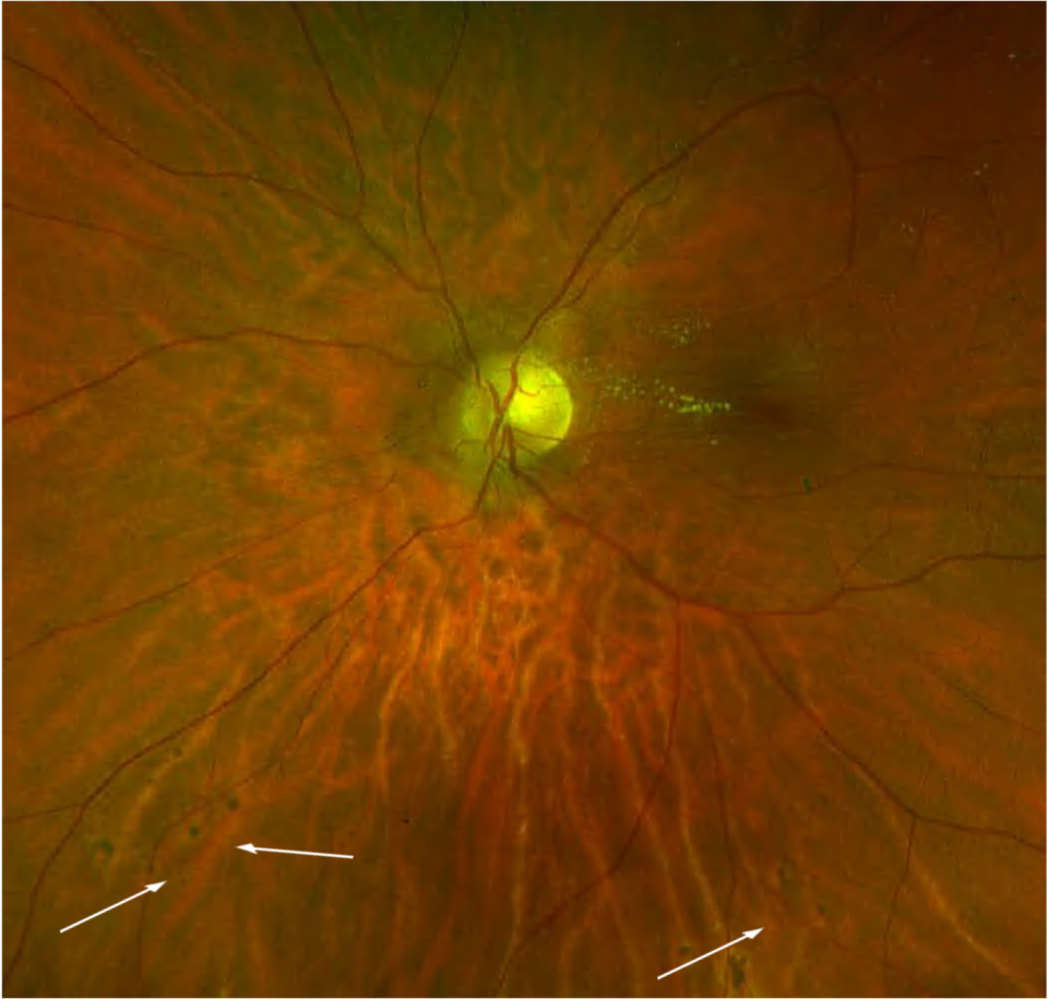
Edited by
Svein Tindlund and Jon Gjelle
Published
June 2023

Sections
01
Introduction

Read more
02
Symptoms

Read more
03
Clinical signs

Read more
04
Diagnostic procedures

Read more
05
Management and treatment

Read more
06
References

Read more
01

Introduction

Hypertension is the leading risk factor for cardiovascular disease (CVD) and mortality worldwide, with a projected number of 1.56 billion individuals with hypertension by 2025.(1,2)

The American College of Cardiology/American Heart Association (ACC/AHA) suggested the following definitions for high blood pressure in 2017.(3)

Category 

Systolic (SBP) 

Diastolic (DBP) 

Elevated blood pressure  

120-129 mmHg 

< 80 mmHg 

Stage 1 hypertension 

130-139 mmHg 

80-89 mmHg 

Stage 2 hypertension 

≥ 140 mmHg 

 ≥ 90 mmHg 

Hypertension profoundly affects the vasculature’s structure and function in the eye. This results in various clinical signs representing hypertensive retinopathy, choroidopathy, and optic neuropathy. Hypertensive retinopathy is the most common clinical presentation. In addition, hypertension may cause occlusion of the main retinal vessels, representing retinal artery or retinal vein occlusions (both branch and central).

Hypertensive retinopathy ranges from 2-17% in non-diabetic patients, but the prevalence varies by demographic group.(4) Hypertensive retinopathy is more common among African, American, and Chinese individuals. Risk factors for essential hypertension include a high salt diet, obesity, tobacco use, alcohol, family history, stress, and ethnic background. -The duration of high blood pressure is the primary risk factor for arteriosclerotic hypertensive retinopathy. In contrast, the degree of blood pressure elevation beyond normal levels is the major risk factor for malignant hypertension.(5)

The initial phase of hypertensive retinopathy is called the vasoconstrictive phase. High blood pressure affects the vascular muscles tonus, which consequently causes constriction of the arterioles (controls the retinal blood flow). Clinically, it represents arteriolar narrowing (focal or generalised). Some studies suggest that arterial narrowing may be a preclinical marker of hypertension, as some patients may not have developed clinical hypertension yet.(6) Persistently elevated blood pressure leads to the sclerotic phase, which causes thickening of the intima of blood vessels and media wall hyperplasia (structural changes of retinal arterioles). This stage accords with diffused and localised (focal) retinal arteriolar narrowing, arteriolar wall opacification (“silver” or “copper wiring”), and compression of the venules by structural changes in the arterioles (arteriovenous “nicking” or “nipping”). Furthermore, chronically elevated blood pressure disrupts the blood-retinal barrier, which is secondary to necrosis of both smooth muscles of retinal vessels and endothelial cells. -. Retinal microaneurysms, haemorrhages, lipid exudates, and cotton wool spots characterise the exudative phase. (7) Very high blood pressure, which occurs rapidly (above 180/120 mmHg), causes signs of malignant hypertensive retinopathy. Clinically, it manifests with signs of the exudative phase, including optic disc swelling and lipid exudates in the macula.

Recent research findings suggest that generalised retinal arteriolar narrowing and arteriovenous nicking are not only associated with current blood pressure levels but also with blood pressure levels measured in the past. These findings suggest that these retinal signs reflect the cumulative effects of longstanding hypertension and serve as persistent markers of chronic hypertensive damage. On the other hand, focal arteriolar narrowing, retinal haemorrhages, microaneurysms, and cotton-wool spots are associated explicitly with concurrently measured blood pressure levels. These retinal signs mirror the effects of short-term blood pressure changes rather than reflecting the cumulative impact of longstanding hypertension. (8)

In addition, studies found that retinal venular widening or dilation is also related to high blood pressure levels and incident hypertension.

Based on the above description of different stages of the disease, hypertensive retinopathy can be classified as follows(11):

  1. No signs of hypertensive retinopathy
  2. Mild: Generalised arteriolar narrowing, focal arteriolar narrowing, arteriovenous nicking, arteriolar wall opacification (silver or copper wiring), or a combination of these signs.
  3. Moderate: Haemorrhages (blot, dot, or flame-shaped), microaneurysms, cotton-wool spots, hard exudates, or a combination of these signs.
  4. Malignant: Signs of moderate retinopathy in combination with optic disc swelling in the presence of severely elevated blood pressure.
02

Symptoms

Most patients are asymptomatic. Those who represent symptoms complain of headache and blurred vision. It is essential to be familiar with the symptoms of hypertension and its complications. Ask patients about symptoms of hypertension such as headache, eye pain, chest pain, shortness of breath, dyspnoea on exertion, palpitations, orthopnea , and paroxysmal nocturnal
dyspnea .(5)

In addition, always ask about blood pressure levels and medication compliance.

Remember that hypertension is related to stroke, ischaemic heart disease, cognitive impairment, and renal impairment. – Recognising the clinical signs of hypertensive retinopathy is vital, as they can often serve as the initial and prominent indicators of high blood pressure.

03

Clinical signs

Clinical signs depend on the stage of hypertensive retinopathy and the chronicity of -high blood pressure.

Acute hypertensive retinopathy can be classified using the modified Scheie Classification of Hypertensive Retinopathy(5):

  • Grade 0: No changes.
  • Grade 1: Barely detectable arterial narrowing.
  • Grade 2: Obvious arterial narrowing with focal irregularities.
  • Grade 3: Grade 2 plus retinal haemorrhages, exudates, cotton wool spots, or retinal oedema.
  • Grade 4: Grade 3 plus optic disc swelling.

The Sheie Classification is used to classify chronic hypertensive retinopathy changes as follows(12)

  • Stage 1: Widening of the arteriole light reflex
  • Stage 2: Stage 1 + Arteriovenous crossing sign
  • Stage 3: Copper wiring of arterioles (copper-coloured arteriole light reflex)
  • Stage 4: Silver wiring of arterioles (silver-coloured arteriole light reflex).
Image 1. Grade 3 hypertensive retinopathy – right eye
Image 2. Grade 3 hypertensive retinopathy – left eye (same patient as compared to Image)
Image 3. Colour fundus photo of a diabetic patient with combined diabetic retinopathy and hypertensive retinopathy. Note that dilated retinal veins and cotton wool spots favour hypertensive retinopathy more than diabetic retinopathy.
Image 4. Lipid exudates and optic disc oedema as part of grade 4 hypertensive retinopathy
04

Diagnostic procedures

Slit lamp examination and colour fundus photography are classical examination methods where it is crucial to be familiar with the clinical signs mentioned above.

Image 5. Signs of chronic hypertensive retinopathy with signs of an acute episode of high blood pressure (lipid exudates). Pigmented dots (Elschnig spots) are signs of hypertension choroidopathy.

Optical coherence tomography (OCT) can be a valuable tool for assessing the macula and detecting the presence of subretinal fluid. It is particularly useful in cases of malignant hypertensive retinopathy, as some presentations may involve subretinal fluid accumulation.

Accurate blood pressure measurement is crucial. It is important to ensure that you are properly trained to measure blood pressure or have access to automated medical devices in your practice. Always measure blood pressure to obtain reliable and precise readings.

05

Management and treatment

Treatment of hypertensive retinopathy is based on the regulation of blood pressure. Trained physicians do it. Eye care specialists monitor retinal changes and treat eye complications like retinal vein occlusion.

Sometimes, non-experienced physicians may misdiagnose malignant hypertension with central retinal vein occlusion and initiate treatment with intravitreal agents. Remember that malignant hypertension is always bilateral and relatively symmetrical. Central retinal vein occlusion is rarely bilateral and symmetrical.

06

References

1 Lawes CM, Vander Hoorn S, Rodgers A. Global burden of blood-pressure-related disease, 2001. The Lancet. 2008 May 3;371(9623):1513-8.

2 Kearney PM, Whelton M, Reynolds K, Muntner P, Whelton PK, He J. Global burden of hypertension: analysis of worldwide data. The lancet. 2005 Jan 15;365(9455):217-23.

3 2017 ACC/AHA/AAPA/ABC/AAPA/AGS/APhA/ASH/ASPC/NMA/PCNA Guideline for the Prevention, Detection, Evaluation, and Management of High Blood Pressure in Adults: A Report of the American College of Cardiology/American Heart Association Task Force on Clinical Practice Guidelines.

4 Nwankwo T, Yoon SS, Burt VL. Hypertension among adults in the United States: national health and nutrition examination survey, 2011-2012. US Department of Health and Human Services, Centers for Disease Control and Prevention, National Center for Health Statistics; 2013.

5 https://eyewiki.aao.org/Hypertensive_Retinopathy#cite_note-2

6 Ding J, Wai KL, McGeechan K, Kawasaki R, Xie J, Klein R, Klein BB, Cotch MF, Wang JJ, Mitchell P, Shaw JE. Retinal vascular caliber and the development of hypertension: a meta-analysis of individual participant data. Journal of hypertension. 2014 Feb;32(2):207.

7 C Y Cheung, Tien Y Wong. HypertensionIn: Schachat AP, Wilkinson CP, Hinton DR, Sadda SR, Wiedemann P, eds. Ryan’s Retina. 6th ed.Philadelphia. Elsevier/Saunders; 2018:chap52.

8 Wong TY, Hubbard LD, Klein R, Marino EK, Kronmal R, Sharrett AR, Siscovick DS, Burke G, Tielsch JM. Retinal microvascular abnormalities and blood pressure in older people: the Cardiovascular Health Study. British Journal of Ophthalmology. 2002 Sep 1;86(9):1007-13.

9 Sun C, Wang JJ, Mackey DA, Wong TY. Retinal vascular caliber: systemic, environmental, and genetic associations. Survey of ophthalmology. 2009 Jan 1;54(1):74-95.

10 Ding J, Wai KL, McGeechan K, Kawasaki R, Xie J, Klein R, Klein BB, Cotch MF, Wang JJ, Mitchell P, Shaw JE. Retinal vascular caliber and the development of hypertension: a meta-analysis of individual participant data. Journal of hypertension. 2014 Feb;32(2):207.

11 Downie LE, Hodgson LA, DSylva C, McIntosh RL, Rogers SL, Connell P, Wong TY. Hypertensive retinopathy: comparing the Keith–Wagener–Barker to a simplified classification. Journal of hypertension. 2013 May 1;31(5):960-5.

12 Good WV. Ophthalmology: A Pocket Textbook Atlas.