Intermediate uveitis

Referral priority: Moderate or urgent

Click on one of the cards below to read more about the specific eye condition.

Written by
Marko Lukic
Edited by
Svein Tindlund and Jon Gjelle
Published
June 2023

Sections
01
Introduction

Read more
02
Symptoms

Read more
03
Clinical features

Read more
04
Diagnostic procedures

Read more
05
Management and treatment

Read more
06
References

Read more
01

Introduction

Uveitis is an inflammation of the uveal tract and can be divided into anterior (iris and ciliary body) and posterior components (choroid). The inflammation may affect surrounding tissue like the retina and optic nerve.(1) Uveitis – particularly posterior uveitis –is a common cause of preventable blindness, and is thus deemed a sight-threatening condition. It is responsible for between 10% to 15% of all cases of blindness and 30,000 new cases of legal blindness annually in the United States.(2)(3) In Western countries, uveitis affects 200 per 100,000 people.(4) More than 35% of patients who develop the condition suffer severe visual impairment.(4,5)

There are four types of uveitis, classified by anatomical location:

Type of uveitis  Primary location 
Anterior  Iris, ciliary body, or both 
Intermediate uveitis  Vitreous 
Posterior uveitis  Choroid 
Panuveitis  The entire uveal tract and vitreous 


Then, uveitis is further classified based on the following characteristics:(6)

  • Onset (sudden versus insidious)
  • Duration (limited, less than 3 months; persistent, more than 3 months)
  • Course (acute, recurrent, or chronic)
  • Laterality (unilateral versus Bilateral)

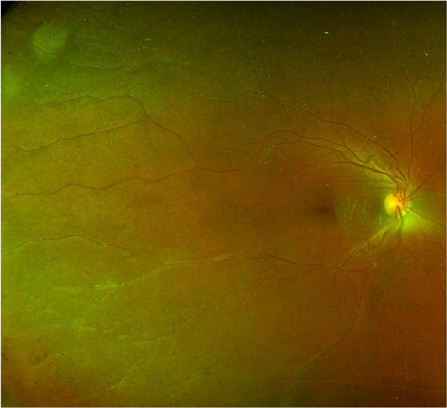
Intermediate uveitis is an inflammation of the vitreous and peripheral retina.(7) It is a chronic, relapsing disease with insidious onset.(8) Patients with intermediate uveitis may have changes in the peripheral retina (peripheral vasculitis) and/or macular oedema. Those features do not change the classification of uveitis (as agreed in the SUN working group).(7) In literature, we use different terms for the same or similar clinical features: intermediate uveitis (IU), pars planitis, chronic cyclitis, peripheral uveitis, vitritis, cyclochorioretinitis, chronic posterior cyclitis, and peripheral uveoretinitis.9 However, we use the term pars planitis when clinically in the presence of snow banks/balls without infectious or systemic cause (SUN classification). We use the term intermediate uveitis when there is presence of infectious or systemic causes.(7)

The percentage of intermediate uveitis varies depending on geographic location. In Western countries, it has been reported in 1.4-22% of all uveitis cases with a prevalence of 5.9/100,000.(9-14) In India, intermediate uveitis is present in 9.5-17.4%.(15-17) Intermediate uveitis accounts for 10 to 12% of all uveitis in children.(18) The idiopathic (unknown cause – termed as pars planitis) form of intermediate uveitis is present in 70% of cases.(19) Other causes may be infectious (tuberculosis, leprosy, Lyme’s disease, syphilis, Toxocara, Whipple’s disease) and non-infectious (sarcoidosis, multiple sclerosis, inflammatory bowel disease, lymphoma, tubulointerstitial nephritis and uveitis syndrome (TINU)).(19,20) Be aware that about 3 to 27% of patients with multiple sclerosis (MS) develop intermediate uveitis/pars planitis, and 7.8 to 14.8% of patients with intermediate uveitis/pars planitis develop MS.(21-23)

Intermediate uveitis typically affects people between 15-40 years. It is bilateral but asymmetrical. The incidence is equal in both men and women.

The condition is initiated by an unknown antigen or may be an autoimmune response. It is mediated by CD+4 T lymphocytes.(24,25) There is also an association with HLA genotypes (HLA-DR, B8, and B51), the most significant being HLA-DR which occurs in 67 to 72% of patients.(26,27)

02

Symptoms

The typical symptoms of intermediate uveitis are blurry vision and floaters. Redness, pain in the eye, and photophobia are rare symptoms compared to acute anterior uveitis cases. Bilateral involvement at initial presentation approaches 80%, although it is frequently asymmetric. Eventually, approximately one-third of unilateral cases will become bilateral.

It is critical to take a thorough medical history of your patient, including present and past personal medical history, surgeries, history of trauma, medicines, and family history. Always ask your patient about arthritis, rashes, shortness of breath, swollen lymph nodes, recent headaches, hearing difficulties, hair loss, pigment changes in the skin, a history of ocular trauma, recent insect bites, sexually transmitted diseases (STDs), tuberculosis (TB) exposure, blood in stools, and recent travels.(29-31)

03

Clinical features

The clinical findings of intermediate uveitis depend on the cause. Up to 50% of cases may have mild to moderate inflammation of anterior eye segments, which includes keratitis precipitates and cells in the anterior chamber. Those findings are most often in children and patients with MS.

Keratitic precipitates (KPs) are white blood cells present on the corneal endothelium. Small, white KPs are a sign of the non-granulomatous type of inflammation, while large, yellowish to brown KPs are significant for the granulomatous type of uveitis.

Competence in examining the anterior chamber is crucial in recognising and monitoring anterior uveitis. Flares and cells in the anterior chamber are signs of intraocular inflammation. Therefore, the cells in the anterior chamber should be graded by severity under high-magnification slit lamp examination in a 1 X 1-mm field of light, as described by the SUN Working Group Grading Scheme for Anterior Chamber Cells, as follows:(32)

  • 0 < 1 cell
  • 0.5 = 1-5 cells
  • 1+ = 6-15 cells
  • 2+ = 16-25 cells
  • 3+ = 26-50 cells
  • 4+ = More than 50 cells

Flare is the presence of proteins in the anterior chamber, and the SUN Working Group proposed the below classification:(15)

  • 0 = None
  • 1+ = Faint
  • 2+ = Moderate (iris and lens details clear)
  • 3+ = Marked (iris and lens details hazy)
  • 4+ = Intense (fibrin or plastic aqueous)

Vitreous cells are universal findings in patients with intermediate uveitis. Mostly, they are present in the anterior vitreous. The counting of cells is used, using the same principle as mentioned above for the cells in the anterior chamber.

Vitreous haze may be present in severe cases. The SUN classification is as below (it may be very subjective):(33)

  • Score 0 – No clinical findings
  • Score 1 – Posterior pole clearly visible
  • Score 2 – Posterior pole details hazy
  • Score 3 – Posterior pole very hazy
  • Score 4 – Posterior pole barely visible

Snowballs are aggregates of vitreous cells, mostly located in the inferior vitreous. They are usually white or yellow. They are typical findings in intermediate uveitis, particularly in pars planitis.

Snowbanks are exudates present as plaques in inferior ora serrata. Scleral indentation or indirect ophthalmoscopy is sometimes required to srecognise those findings.

Peripheral periphlebitis is inflammation of peripheral venules and may be present in the form of sheathing. It may appear months or years after the initial presentation.

Complications of intermediate uveitis may be early or late:

  • Early: Cystoid macular oedema (28-25% of cases), Optic disc oedema (most often present in children younger than 16), synechiae. Be aware that patients with 6/6 vision may also have the presence of macular oedema.
  • Late: Posterior subcapsular cataract, glaucoma, new vessels of the peripheral retina, and retinal ischaemia. Vitreous haemorrhage is most often present in children. Retinal detachments are also described. In addition, cyclitic membranes (fibrovascular tissue) may develop on the ciliary body, which causes hypotony and phthisis of the bulbus. Band keratopathy (corneal calcifications) is one of the late complications as well.
04

Diagnostic procedures

Slit lamp examination is crucial in the examination of patients with anterior uveitis.

Colour fundus photos are useful in checking posterior eye segments and recognising signs of intermediate and posterior uveitis.

Optical coherence tomography is a golden standard in diagnosing uveitis macular oedema. Intermediate uveitis may be complicated with macular oedema, and OCT scans are important in the workup with uveitis patients.

Ultrasound biomicroscopy can be used to evaluate patients with intermediate uveitis. It uses higher frequencies than ocular ultrasound (35-100 MHz) and provides high-resolution images of the pars plana region. It can detect snow banking, cyclitic membranes, vitreous membranes, and peripheral vitreoretinal tractions.

Fundus fluorescein angiography is useful to diagnose and monitor cases of intermediate uveitis.

Image 1. Case of sarcoidosis. You can see peripheral vasculitis (called periphlebitis as retinal veins are inflamed) in the image.
Image 2. Cystoid macular oedema in case of intermediate uveitis secondary to sarcoidosis.
05

Management and treatment

While mild asymptomatic vitreous cells may not require immediate treatment, any evidence of cystoid macular oedema (CME), neovascularisation of the peripheral retina, extensive vasculitis, or significant vitreous cell requires treatment.

Malignancy and infection must be ruled out before starting non-specific anti-inflammatory therapy.

Treatment of uveitis should be prescribed by an experienced eye care specialist.

Steroids are the most important part of treatment. Local steroid eye drops are used to control the inflammation of the anterior segments (if there is the presence of keratitic precipitates (KPs) and/or cells in the anterior chamber).

Unilateral cases are mostly treated with periorbital steroid injections of Triamcinolone. If they have no effect or if there is bilateral involvement, at the same time, oral prednisolone is the treatment of choice. A purified protein derivative (PPD) test is imperative before starting any patient on systemic corticosteroids if there are any risk factors for tuberculosis (TB). Once the inflammation stabilises, you can taper the oral dose according to disease activity. An H2 blocker or a proton pump inhibitor can be prescribed adjunctively to oral steroids (to protect the stomach). Reluctant cases or those with CME require intravitreal treatment with steroids. The treatment of choice is intravitreal Triamcinolone, intravitreal Dexamethasone implant, and intravitreal implant of Fluocinolone acetonide.

If corticosteroids cannot control the intermediate uveitis or in patients whose disease invariably flares when steroids are stopped, immunosuppressive therapy often is attempted. Immunosuppression or immune modulation is also used as part of steroid-sparing therapy to reduce the ’patient’s requirement for systemic corticosteroids and, therefore, to diminish the adverse effects of systemic corticosteroid therapy.

06

References

1 Dunn JP. Uveitis. Prim Care. 2015 Sep. 42 (3):305-23.

2 Nussenblatt RB. The natural history of uveitis. International ophthalmology. 1990 Oct;14(5):303-8.

3 DARRELL RW, WAGENER HP, KURLAND LT. Epidemiology of uveitis: incidence and prevalence in a small urban community. Archives of ophthalmology. 1962 Oct 1;68(4):502-14.

4 London NJ, Rathinam SR, Cunningham ET. The epidemiology of uveitis in developing countries. International Ophthalmology Clinics. 2010 Apr 1;50(2):1-7.

5 Rothova A, Suttorp-van Schulten MS, Frits Treffers W, et al. Causes and frequency of blindness in patients with intraocular inflammatory disease. Br J Ophthalmol. 1996;80:332–336.

6 Rao NA. Uveitis and other intraocular inflammations. Ophthalmology. 2nd ed. Philadelphia: Mosby. 2004:1105-238.

7 Standardization of Uveitis Nomenclature (SUN) Working Group. Standardization of uveitis nomenclature for reporting clinical data. Results of the First International Workshop. American journal of ophthalmology. 2005 Sep 1;140(3):509-16.

8 Brad Bowling. Kanski’s Clinical Ophthalmology – A Systematic Approach, 8th edition. Elsevier , 2016.

9 Vitale AT, Zierhut M, Foster CS. Intermediate uveitis. Diagnosis and treatment of uveitis. Philadelphia: WB Saunders and Company. 2002:844-57.

10 McCANNEL CA, Holland GN, Helm CJ, Cornell PJ, Winston JV, Rimmer TG, UCLA Community-Based Uveitis Study Group. Causes of uveitis in the general practice of ophthalmology. American journal of ophthalmology. 1996 Jan 1;121(1):35-46.

11 Chan SM, Hudson M, Weis E. Anterior and intermediate uveitis cases referred to a tertiary centre in Alberta. Canadian Journal of Ophthalmology. 2007 Dec 1;42(6):860-4.

12 Henderly DE, Genstler AJ, Smith RE, Rao NA. Changing patterns of uveitis. American journal of ophthalmology. 1987 Feb 1;103(2):131-6.

13 Rodriguez A, Calonge M, Pedroza-Seres M, Akova YA, Messmer EM, D’Amico DJ, Foster CS. Referral patterns of uveitis in a tertiary eye care center. Archives of ophthalmology. 1996 May 1;114(5):593-9.

14 Vadot E. Epidemiology of intermediate uveitis: a prospective study in Savoy. Intermediate Uveitis. 1992;23:33-4.

15 Singh R, Gupta V, Gupta A. Pattern of uveitis in a referral eye clinic in north India. Indian journal of ophthalmology. 2004 Apr 1;52(2):121-5.

16 Das D, Bhattacharjee H, Das K, Tahiliani PS, Bhattacharyya P, Bharali G, Das M, Deka A, Paul R. The changing patterns of uveitis in a tertiary institute of Northeast India. Indian journal of ophthalmology. 2015 Sep 1;63(9):735-7.

17 Rathinam SR, Namperumalsamy P. Global variation and pattern changes in epidemiology of uveitis. Indian journal of ophthalmology. 2007 May 1;55(3):173-83.

18 Engelmann K, Ness T, Greiner K, Hudde T. Uveitis intermedia in childhood. Klinische Monatsblatter fur Augenheilkunde. 2007 Jun 1;224(6):462-8.

19 https://eyewiki.aao.org/Intermediate_Uveitis

20 Babu BM, Rathinam SR. Intermediate uveitis. Indian journal of ophthalmology. 2010 Jan 1;58(1):21-7.

21 Porter RI. Uveitis in association with multiple sclerosis. The British Journal of Ophthalmology. 1972 Jun;56(6):478.

22 Breger BC, Leopold IH. The incidence of uveitis in multiple sclerosis. American journal of ophthalmology. 1966 Sep 1;62(3):540-5.

23 Chester GH, Blach RK, Cleary PE. Inflammation in the region of the vitreous base. Pars planitis. Transactions of the Ophthalmological Societies of the United Kingdom. 1976 Apr 1;96(1):151-7.

24 Wetzig RP, Chan CC, Nussenblatt RB, Palestine AG, Mazur DO, Mittal KK. Clinical and immunopathological studies of pars planitis in a family. British journal of ophthalmology. 1988 Jan 1;72(1):5-10.

25 Nölle B, Eckardt C. Cellular phenotype of vitreous cells in intermediate uveitis. InIntermediate Uveitis 1992 (Vol. 23, pp. 145-149). Karger Publishers.

26 Tang WM, Pulido JS, Eckels DD, Han DP, Mieler WF, Pierce K. The association of HLA-DR15 and intermediate uveitis. American journal of ophthalmology. 1997 Jan 1;123(1):70-5.

27 Boyd SR, Young S, Lightman S. Immunopathology of the noninfectious posterior and intermediate uveitides. Survey of ophthalmology. 2001 Nov 1;46(3):209-33.

28 https://emedicine.medscape.com/article/1208794-clinical

29 Sanghvi C, Bell C, Woodhead M, Hardy C, Jones N. Presumed tuberculous uveitis: diagnosis, management, and outcome. Eye. 2011 Apr;25(4):475-80.

30 Kamal S, Kumar R, Kumar S, Goel R. Bilateral interstitial keratitis and granulomatous uveitis of tubercular origin. Eye & Contact Lens. 2014 Mar 1;40(2):e13-5.

31 Abderrahim K, Chebil A, Falfoul Y, Bouladi M, Matri LE. Granulomatous uveitis and reactive arthritis as manifestations of post-streptococcal syndrome. International ophthalmology. 2015 Oct;35(5):641-3.

32 Standardization of Uveitis Nomenclature (SUN) Working Group. Standardization of uveitis nomenclature for reporting clinical data. Results of the First International Workshop. American journal of ophthalmology. 2005 Sep 1;140(3):509-16.

33 Davis JL, Madow B, Cornett J, Stratton R, Hess D, Porciatti V, Feuer WJ. Scale for photographic grading of vitreous haze in uveitis. American journal of ophthalmology. 2010 Nov 1;150(5):637-41.