Keratoconus and corneal
ectasia

Referral priority:  Routine or urgent 

All patients with active hydrops or corneal rupture must be urgently referred to an ophthalmologist following local guidelines. Consider referral for corneal cross-linking in recently diagnosed cases and progressing cases. Management with contact lenses should be undertaken by a specialised optometrist with experience in managing and fitting keratoconus.

Click on one of the cards below to read more about the specific eye condition.

Written by
Marko Lukic
Edited by
Svein Tindlund and Jon Gjelle
Published
June 2023

Sections
01
Introduction

Read more
02
Symptoms

Read more
03
Clinical signs

Read more
04
Diagnostic procedures

Read more
05
Management and treatment

Read more
06
References

Read more
01

Introduction

Ectatic corneal disease (ECD) comprises a group of disorders characterised by progressive thinning and subsequent bulging of the corneal structure.(1,2) There are different phenotypes described in the literature: keratoconus (KC), keratoglobus (KG), and pellucid marginal degeneration (PMD).(1) Furthermore, progressive iatrogenic corneal ectasia has been reported after different refractive laser procedures and has been grouped within the ectatic corneal disease.(3,4) Throughout time, various names for the above-named conditions were used. In 2015, the Global consensus published recommendations for diagnosing and managing corneal ectatic diseases.(5)

Keratoconus (KC)
Keratoconus is a bilateral, typically asymmetrical, corneal disorder where the central or paracentral cornea undergoes progressive thinning and steeping, causing irregular astigmatism and blurred vision.(2,6) The aetiology of the disease is unknown. However, it is associated with several conditions like atopy, Down’s Syndrome, Leber’s congenital amaurosis, and Brittle cornea syndrome/Ehler’s Danlos/connective disorders. The reported prevalence is 1 in 700.(7) There are some risk factors associated with keratoconus: eye rubbing (atopy and vernal keratoconjunctivitis), Down’s Syndrome, sleep apnoea, connective tissue disorders, floppy eyelid syndrome, positive family history, and retinitis pigmentosa.(8) Signs of keratoconus, depending on its severity, are breaks in Bowman’s membrane, collagen disorganisation, scaring, and corneal thinning.  

Keratoglobus (KG)
Keratoglobus is characterised by a generalised thinning and globular protrusion of the cornea, which results in irregular corneal topography with increased corneal fragility due to extreme thinning.(9) There are two major forms, congenital and acquired form. The congenital one is associated with blue sclera syndrome, Leber congenital amaurosis, and Brittle cornea syndrome (previously described as Ehler-Danlos type VIB).(18) The acquired form evolves from keratoconus and Pellucid Marginal Degeneration (PMD), and is associated with vernal keratoconjunctivitis, dysthyroid orbitopathy, and chronic marginal blepharitis.(10) The aetiology is unknown, and risk factors are not identified. There is the presence of diffuse corneal thinning and focal breaks of Bowman’s membrane, which are most severe in the peripheral cornea.

Pellucid Marginal Degeneration (PMD)
Pellucid Marginal Degeneration (PMD) is a non-inflammatory, nonhereditary cause of corneal ectasia with bilateral, clear, inferior (typically 4 o’clock to 8 o’clock), peripheral corneal thinning. It is characterised by a crescent-shaped band of inferior corneal thinning approaching 20% of normal thickness that is 1 to 2 mm in height, 6 to 8 mm in horizontal extent, and 1 to 2 mm from the limbus. There is no associated inflammation, and the central cornea is of normal thickness.(8,11) Generally, it is rare but the second most common corneal ectasia. Ten per cent of PMD cases are associated with keratoconus, and 13% are associated with keratoglobus.(8) Histological findings are similar to those found in keratoconus.

Ectasia
Ectasia is a rare complication of corneal refractive surgery, and occurs in just 0.04% to 0.6% of procedures.(12) A large study found that 96% of ectasia cases occurred due to Laser-Assisted In Situ Keratomileusis (LASIK) and 4% due to Photorefractive keratectomy (PRK).(13) There are also cases of ectasia reported after the Small incision lenticule extraction (SMILE) procedure.(14) It results from a loss of biomechanical integrity of the cornea with subsequent thinning and steepening of the tissue. Screening for cases at higher risk for ectasia progression became a significant concern in the preoperative phase of elective refractive procedures.(15)

02

Symptoms

Keratoconus is a bilateral condition, but in most cases, asymmetrical. It typically occurs in early adolescence and progresses in the 20s or 30s. The less affected eye may show a high amount of astigmatism or mild steepening. There is often a history of frequent changes in glasses prescription that do not adequately correct vision. Patients manifest with high or progressive astigmatism, which may be an early sign of the condition. More advanced cases manifest with blurry and decreased vision.

Patients with keratoglobus represent a bilateral decrease in visual acuity, which cannot be improved with spectacles or contact lenses. Those who develop hydrops (stromal edema due to leakage of aqueous humor through a tear in Descemet’s membrane) or corneal rupture are represented with eye pain.

Patients with PMD normally present asymptomatically, but cases can also present with progressive visual deterioration despite spectacle correction due to irregular astigmatism.

Patients who underwent laser refractive surgery at a younger age might have an increased risk for ectasia.

03

Clinical signs

There are early and late clinical signs which indicate keratoconus.(8)

Early signs

  • Scissoring of the light reflex on retinoscopy: occurs due to high irregular corneal astigmatism.
  • Corneal thinning on a slit lamp, particularly in the inferior cornea.
Image 1. Inferior corneal thinning in a patient with pellucid marginal degeneration.
  • Rizzuti’s sign: conical reflection on the nasal cornea when a penlight is shone from the temporal side.
  • Fleischer ring: an iron deposit often present within the epithelium around the base of the cone. It is brown and best visualised with a cobalt blue filter.
  • Vogt’s striae: fine, roughly vertically parallel striations in the stroma. These generally disappear with firm pressure applied over the eyeball and re-appear when pressure is stopped.

Late signs

  • Munson’s sign: a protrusion of the lower eyelid in downgaze.
  • Breaks in Bowman’s membrane.
  • Acute hydrops: a condition where a break in the Descemet’s membrane allows aqueous to enter into the stoma, causing severe corneal thickening, decreased vision, light sensitivity, tearing, and pain.
  • Stromal scarring after the resolution of acute hydrops, which paradoxically may improve vision in some cases by changing corneal curvature and reducing irregular astigmatism.

Patients with keratoglobus represent the globular corneal appearance and diffuse corneal thinning, mostly in the peripheral retina. They may also develop hydrops and corneal scarring.

Image 2. Hydrops in a patient with keratoconus. Corneal haze and increased corneal oedema. Mild redness of the eye.

Patients with PMD manifest inferior corneal thinning. Their visual acuity cannot be improved with spherocylindrical lenses. However, it can be improved with a pinhole. The steepest corneal protrusion in PMD occurs above (central to) the area of stromal thinning, appearing like a ”beer belly” in the cross-section. This results in high and irregular ”against-the-rule” astigmatism of up to 20 diopters and a flattening of the vertical meridian. Astigmatism can instead be ”with the rule” if the inferior curvature is steeper than the horizontal curvature. The thinning area in PMD is always epithelialised, clear, avascular, and without lipid deposition (distinguishing it from Terrien marginal degeneration).

04

Diagnostic procedures

Slit lamp examination is the first step to diagnose corneal ectasia (mostly moderate and advanced stages), following the specific signs and checking corneal thickness using light beams.

Pachymetry is the measurement of corneal thickness, which can be done in several ways (i.e., using anterior OCT). It is very useful as additional proof of corneal thinning after slit lamp examination.

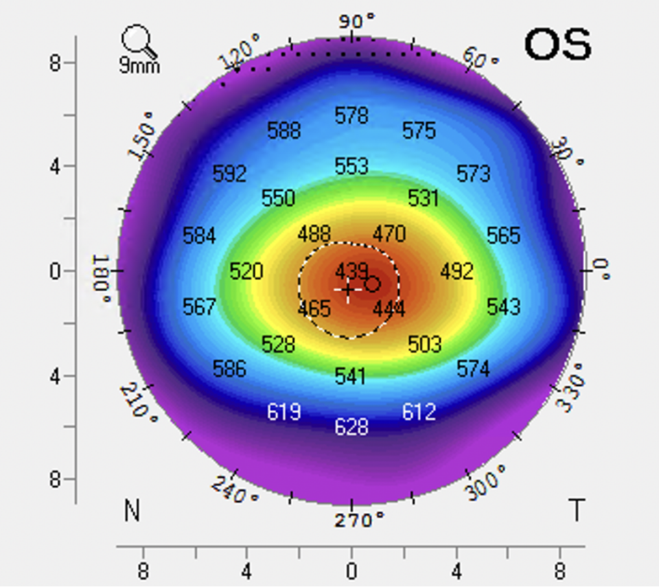
Computerised corneal topography is a computer-assisted diagnostic imaging technique in which a special instrument projects a series of light rings on the cornea, creating a colour-coded map of the corneal surface and cross-section profile.(16) Those medical devices produce a 3D image that characterises anterior and posterior corneal curvature and thickness distribution. They provide data on central corneal thickness, thinnest corneal thickness, changes in corneal thickness over the entire cornea, anterior curvature radius, and posterior curvature radius. It is crucial in preoperative refractive surgery diagnosis. It recognises early forms of corneal ectasia and monitors post-refractive surgery patients

Image 3. Computerised corneal topography image of the left eye. Corneal thinning in the central zone in a patient with keratoconus.

Refractive error must be considered in conjunction with corneal thickness to determine if the patient will have an adequate residual stromal bed thickness. Approximately 15 µm of stromal tissue is ablated for every diopter of myopia correction, and a low residual stromal bed thickness (250–300 µm) increases the risk for postoperative ectasia.(17)

05

Management and treatment

Some mild cases of keratoconus can be treated with soft toric lenses. However, most cases require rigid gas-permeable contact lenses or scleral lenses to neutralise irregular astigmatism.

The primary treatment for progressive keratoconus is corneal cross-linking. Corneal collagen cross-linking is a minimally invasive treatment using riboflavin and UV light to induce corneal stroma stiffening by forming additional cross-link bonds within the extracellular matrix of the stromal collagen. There are different protocols used. Nonetheless, the conventional epithelium-off procedure showed a high success rate. Patients are monitored every 3-6 months.

Patients who develop active hydrops require cycloplegics to relieve pain and hypertonic eye drops/ointment (sodium chloride 5%).

In most advanced cases, when corneal cross-linking does not provide satisfactory results, surgical options may be considered (intrastromal corneal ring segments (ISCR), deep anterior lamellar keratoplasty (DALK), or penetrating keratoplasty (PK).

06

References

1 Salomao MQ, Hofling-Lima AL, Esporcatte LP, Correa FF, Lopes B, Sena Jr N, Dawson DG, Ambrosio Jr R. Ectatic diseases. Experimental Eye Research. 2021 Jan 1;202:108347.

2 Krachmer JH, Feder RS, Belin MW. Keratoconus and related noninflammatory corneal thinning disorders. Survey of ophthalmology. 1984 Jan 1;28(4):293-322.

3 Seiler T, Koufala K, Richter G. Iatrogenic keratectasia after laser in situ keratomileusis. Journal of refractive surgery. 1998 May 1;14(3):312-7.

4 Seiler T, Quurke AW. Iatrogenic keratectasia after LASIK in a case of forme fruste keratoconus. Journal of Cataract & Refractive Surgery. 1998 Jul 1;24(7):1007-9.

5 Gomes JA, Tan D, Rapuano CJ, Belin MW, Ambrósio Jr R, Guell JL, Malecaze F, Nishida K, Sangwan VS. Global consensus on keratoconus and ectatic diseases. Cornea. 2015 Apr 1;34(4):359-69.

6 Roberts CJ, Dupps Jr WJ. Biomechanics of corneal ectasia and biomechanical treatments. Journal of Cataract & Refractive Surgery. 2014 Jun 1;40(6):991-8.

7 Hashemi H, Heydarian S, Hooshmand E, Saatchi M, Yekta A, Aghamirsalim M, Valadkhan M, Mortazavi M, Hashemi A, Khabazkhoob M. The prevalence and risk factors for keratoconus: a systematic review and meta-analysis. Cornea. 2020 Feb 1;39(2):263-70.

8 https://eyewiki.aao.org/Keratoconus#cite_note-1

9 Wallang BS, Das S. Keratoglobus. Eye. 2013 Sep;27(9):1004-12.

10 Meghpara B, Nakamura H, Vemuganti GK, Murthy SI, Sugar J, Yue BY, Edward DP. Histopathologic and immunohistochemical studies of keratoglobus. Archives of ophthalmology. 2009 Aug 1;127(8):1029-35.

11 Martínez-Abad A, Piñero DP. Pellucid marginal degeneration: Detection, discrimination from other corneal ectatic disorders and progression. Contact Lens and Anterior Eye. 2019 Aug 1;42(4):341-9.

12 Wolle MA, Randleman JB, Woodward MA. Complications of refractive surgery: ectasia after refractive surgery. International ophthalmology clinics. 2016;56(2):129.

13 Randleman JB, Woodward M, Lynn MJ, Stulting RD. Risk assessment for ectasia after corneal refractive surgery. Ophthalmology. 2008 Jan 1;115(1):37-50.

14 Moshirfar M, Albarracin JC, Desautels JD, Birdsong OC, Linn SH, Hoopes Sr PC. Ectasia following small-incision lenticule extraction (SMILE): a review of the literature. Clinical Ophthalmology (Auckland, NZ). 2017;11:1683.

15 Ambrósio Jr R, Randleman JB. Screening for ectasia risk: what are we screening for and how should we screen for it?. Journal of Refractive Surgery. 2013 Apr 1;29(4):230-2.

16 https://www.cms.gov/medicare-coverage-database/view/lcd.aspx?lcdid=33810&ver=15&bc=CAAAAAAAAAAA

17 Belin MW, Ambrósio Jr R. Corneal ectasia risk score: statistical validity and clinical relevance. Journal of refractive surgery. 2010 Apr 1;26(4):238-40.

18 Burkitt Wright EM, Porter LF, Spencer HL, Clayton-Smith J, Au L, Munier FL, Smithson S, Suri M, Rohrbach M, Manson FD, Black G. Brittle cornea syndrome: recognition, molecular diagnosis and management. Orphanet journal of rare diseases. 2013 Dec;8(1):1-1.