Neovascular age-related macular degeneration

Referral priority: urgent

All patients with suspected neovascular age-related macular degeneration must be urgently referred to an ophthalmologist following local guidelines.

Written by
Marko Lukic
Edited by
Svein Tindlund and Jon Gjelle
Published
June 2023

Sections
01
Introduction

Read more
02
Symptoms

Read more
03
Clinical signs

Read more
04
Diagnostic procedures

Read more
05
Management and treatment

Read more
06
References

Read more
01

Introduction

Age-related macular degeneration (AMD) is a complex disease with genetic and environmental aetiology and is the leading cause of irreversible blindness.(1,2) AMD is the macula’s generative disease, and is characterised by a loss of visual acuity caused by degeneration of choriocapillaris, retinal pigment epithelium (RPE), and photoreceptors.(3,4)

AMD is the leading cause of irreversible vision loss in developed countries in people of 50 years or older. The prevalence increases with age, and is 1.6% in those older than 55 years and 13% in those older than 84 years.(4) The predicted number of patients diagnosed with AMD has significantly grown, and the expected number of those diagnosed with AMD will be 288 million in 2040.(5) It has been calculated that 10-20% of non-exudative cases switch to the exudative form of the disease.(6)

The aetiology of AMD is multifactorial and involves an interplay of genetic, environmental, metabolic, and functional factors, including ageing, family history, smoking, high blood pressure, obesity, hypercholesterolemia, and arteriosclerosis.(7,8) The studies revealed that patients with no signs of AMD are at high risk of developing the disease, from 10-19.2%.(8,9) It is important to look at the correlation between risk factors rather than to monitor them individually. There has been significant progress in understanding the role of genes in the pathophysiology of the disease.

The hallmark of the disease is the presence of drusen. Drusen are focal deposits of extracellular debris that typically form between the basal lamina of the RPE and the inner collagenous layer of the ’Bruch’s membrane. The decreased function of RPE cells and changes in the permeability of Bruch’s membrane led to drusen formation. Furthermore, local inflammation (alternative complement cascade) plays a significant role, which means that it is an important factor in the pathomechanism of the disease.(10) Production of vascular endothelial growth factor (VEGF) has a central role in the formation of the complex of choroidal new vessels (CNV), which is the hallmark of the exudative form of the disease.(11) If the condition remains untreated, patients develop a central disciform scar, which causes irreversible vision loss. It is also essential to follow the second eye, due to the high chance of developing the exudative form there as well.(12)

It is important to mention the condition called idiopathic choroidal polyps.(13) The prevalence is higher in patients of Asian ancestry.(14) The aetiology of the condition is unclear. But since many AMD studies have included patients with polyps, polyps are considered a subtype of AMD, although they have different features and pathomechanism.(14,15)

02

Symptoms

AMD is a slowly progressive condition, and patients are usually asymptomatic at an early stage. However, in some early cases, patients may complain of distorted vision (metamorphopsia), blurred vision, and central and paracentral scotoma. In addition, patients usually complain they have problems with reading, where some letters or a whole word is missing. As the disease progresses, the symptoms become more apparent.

Patients with an exudative type of the disease complain of a sudden decrease in vision. Remember that patients who develop a more advanced stage of a non-exudative form of the disease (centre-involving geographic atrophy) may also complain of “sudden” deterioration in vision.

Patients with AMD have problems with central vision, while the peripheral vision remains intact (if there is no other concomitant eye condition, like glaucoma). The loss of central visual acuity leads to a reduction in activities of daily living, as well as mobility problems and an increased risk of falls, fractures, and depression in older people.(16)

03

Clinical signs

The exudative form of AMD is considered an advanced stage of the disease. The main characteristic of this subtype is the presence of the choroidal neovascular membrane. Some clinical findings may be suggestive of exudative AMD:

Retinal pigment epithelium detachment (PED)
The PED is clinically seen as a dome-shaped elevation of retinal pigment detachment. It may have been surrounded by atrophy and hyperpigmentation. It is not exclusively a sign of a neovascular form of the disease. In addition, the presence of bleeding or neurosensory detachment (presence of subretinal fluid) suggests a choroidal neovascular complex.(11)

Haemorrhage
There is a likelihood of bleeding from the choroidal neovascular complexes. Clinically, the bleeding can be seen as dark red when it is present in subretinal space (suggestive of classic or type 2 CNV) or dark when it is present below the retinal pigment epithelium (RPE) (suggestive of occult or type 1 CNV or polyps). In some cases, it may be represented as a localised intraretinal haemorrhage in retinal angiomatous proliferation (RAP) lesions (type 3 CNV). Remember that haemorrhage is not necessarily present in all active cases of CNV. However, massive haemorrhages indicate the presence of polyps.

Sometimes, subretinal haemorrhages may be massive, which may end up as vitreous haemorrhages.(17)

Image 1. Colour fundus photo of subretinal haemorrhages suggestive on exudative AMD.

Retinal pigment epithelium tear
RPE dehiscence or tears of the RPE have been described as a complication associated with CNV, often in an eye with a serous or fibrovascular PED.(18) Tears occur at the junction of attached and detached RPE. Possible when PEDs can no longer resist the stretching forces from the fluids from the occult CNV in the sub-RPE space or from contractile forces in the underlying fibrovascular tissue.(18,19)

Disciform scar
The scar represents the end stage of the neovascular form of the disease. It is primarily white or yellow and. may have accompanied the presence of pigment or haemorrhage.

Patients with a neovascular form of the disease may also have the presence of signs of a non-exudative form of the disease, including geographic atrophy.

04

Diagnostic procedures

Amsler grid is a helpful tool to detect macular changes at an early stage. If the practice owns diagnostic imaging modalities, like colour fundus photography or OCT, try to combine the results of the Amsler grid test and the images to understand the meaning of the test better. But remember, there may be false negative results.

Slit lamp examination is a straightforward way to recognise the clinical features of the AMD.

Colour fundus photography is a useful imaging modality to document the macular changes and monitor changes during follow-ups. It is a good tool to recognise macular haemorrhages.

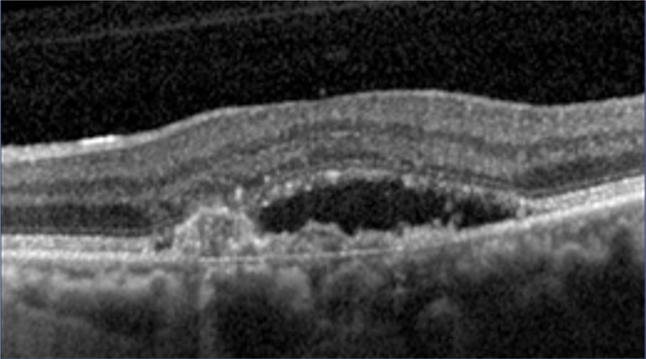
Optical coherence tomography is a golden standard imaging modality to recognise the macular changes related to AMD and to distinguish the entities which may look similar (remember that patients with AMD have drusen). Likewise, it is useful in recognising drusen from pseudodrusen and in identifying signs of exudative AMD or geographic atrophy at an early stage.

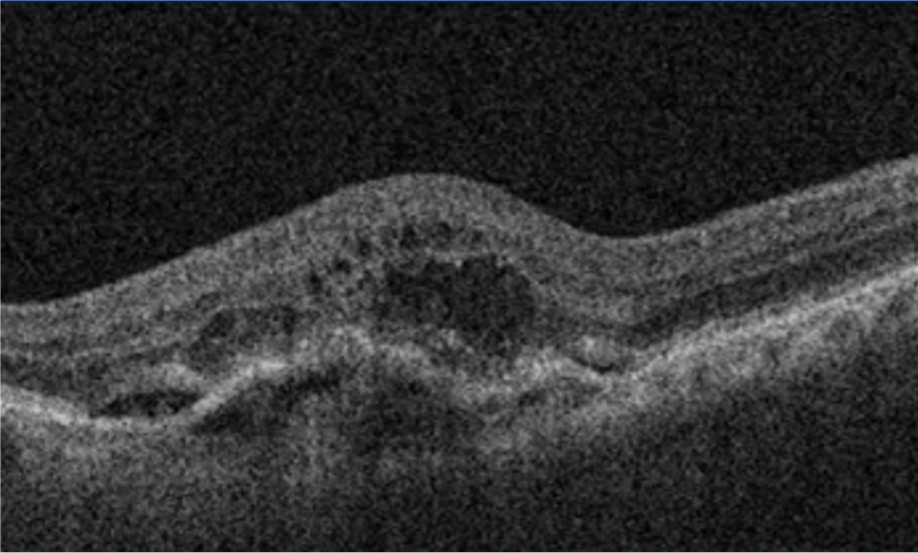
There are three types of CNV based on anatomical location. All three types are present in the neovascular form of the disease. Below are OCT features that describe each type of CNV (20):

  • Type 1 – is located below the retinal pigment epithelium. There is the presence of irregular PED with the presence of subretinal fluid. In literature, based on old terminology, it can be found as an occult membrane.
Image 2. OCT scan of type 1 CNV. Notice irregular PED and subretinal fluid.
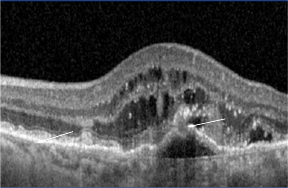
  • Type 2 – is located between the RPE and neurosensory retina. It manifests with the presence of subretinal hyperreflective material and intraretinal fluid. In literature, based on old terminology, it is called classic CNV type.

Likewise, there may be additional features of type 1 (PED and subretinal fluid), which means that the membrane is located both below and above the RPE. In literature, based on the old terminology, it is called mixed CNV.

Image 3. OCT scan of mixed CNV. Notice irregular PED and the presence of intraretinal and subretinal fluid.
  • Type 3 – the neovascular complex starts within the retina, from the deep retinal plexus. Over the progression of the disease, the complex connects with choroidal vessels below the RPE. It manifests as intraretinal hyperreflective material, drusen and intraretinal fluid.
Image 4. OCT scan of active RAP lesion. Irregular PED with intraretinal fluid. The red arrow shows the intraretinal neovascular complex connecting with the PED. The white arrow shows subretinal drusenoid deposits.

Indocyanine green angiography is the most important imaging modality to diagnose polyps. However, nowadays, OCT imaging also is an essential tool to recognise polyps. Some features indicate the active polyps: subretinal fluid with notched pigment epithelium detachments. The PEDs are round or oval structures.(21)

Image 5. OCT scan of polyps. Notice oval-shaped and notched PEDs with the presence of subretinal fluid.

OCT angiography is a novel imaging modality based on the same principles as regular OCT. It reveals the macular perfusion, and is the only one that can distinguish superficial and deep retinal plexuses.(22) It is a non-invasive modality that recognises choroidal neovascular complexes.

05

Management and treatment

Anti-VEGF treatment is the golden standard for the neovascular form of age-related macular degeneration. Multiple randomised and real-life results proved the safety and efficacy of anti-VEGF agents used in clinical protocols: bevacizumab (off-labelled drug), ranibizumab, aflibercept, brolucizumab, and faricimab.(23-35)

Two major treatment protocols are used in long-term management with anti-VEGF: pro re nata (PRN) and treat and extend. PRN uses new injections if new signs of CNV activity are observed at a monthly follow-up. While treat and extend utilises progressive extensions of treatment intervals up to three months depending on clinical findings. Research has shown better improvement in visual acuity with treat and extend compared to PRN.(36)

06

References

1 Friedman DS, O’Colmain BJ, Munoz B, Tomany SC, McCarty C, De Jong PT, Nemesure B, Mitchell P, Kempen J. Prevalence of age-related macular degeneration in the United States. Arch ophthalmol. 2004 Apr 1;122(4):564-72.

2 Sobrin L, Seddon JM. Nature and nurture-genes and environment-predict onset and progression of macular degeneration. Progress in retinal and eye research. 2014 May 1;40:1-5.

3 Rosenfield PJ, Martidis A, Tennant M. Age-related macular degeneration. In: Yanoff M, Duker JS, Augsburger JJ, eds. Ophthalmology: Expert Consult. 3rd ed. Philadelphia, PA: Elsevier Mosby; 2009.

4 Shalev V, Sror M, Goldshtein I, Kokia E, Chodick G. Statin use and the risk of age related macular degeneration in a large health organization in Israel. Ophthalmic epidemiology. 2011 Apr 1;18(2):83-90.

5 Wong WL, Su X, Li X, Cheung CM, Klein R, Cheng CY, Wong TY. Global prevalence of age-related macular degeneration and disease burden projection for 2020 and 2040: a systematic review and meta-analysis. The Lancet Global Health. 2014 Feb 1;2(2):e106-16.

6 Ferris FL, Fine SL, Hyman L. Age-related macular degeneration and blindness due to neovascular maculopathy. Archives of ophthalmology. 1984 Nov 1;102(11):1640-2.

7 Cheung LK, Eaton A. Age‐related macular degeneration. Pharmacotherapy: The Journal of Human Pharmacology and Drug Therapy. 2013 Aug;33(8):838-55.

8 Delcourt C, Souied E, Sanchez A, Bandello F. Development and validation of a risk score for age-related macular degeneration: the STARS Questionnaire. Investigative Ophthalmology & Visual Science. 2017 Dec 1;58(14):6399-407.

9 Bucan K, Lukic M, Bosnar D, Kopic A, Jukic T, Konjevoda S, Glavadanovic S, Gverovic Antunica A. Analysis of association of risk factors for age-related macular degeneration. European Journal of Ophthalmology. 2022 Jan;32(1):410-6.

10 Klein RJ, Zeiss C, Chew EY, Tsai JY, Sackler RS, Haynes C, Henning AK, SanGiovanni JP, Mane SM, Mayne ST, Bracken MB. Complement factor H polymorphism in age-related macular degeneration. Science. 2005 Apr 15;308(5720):385-9.

11 Bressler NM, Bressler SB, Fine SL. Age-related macular degeneration. Survey of ophthalmology. 1988 May 1;32(6):375-413.

12 Thomas MA, Grand MG, Williams DF, Lee CM, Pesin SR, Lowe MA. Surgical management of subfoveal choroidal neovascularization. Ophthalmology. 1992 Jun 1;99(6):952-68.

13 Ciardella AP, Donsoff IM, Huang SJ, Costa DL, Yannuzzi LA. Polypoidal choroidal vasculopathy. Survey of ophthalmology. 2004 Jan 1;49(1):25-37.

14 Imamura Y, Engelbert M, Iida T, Freund KB, Yannuzzi LA. Polypoidal choroidal vasculopathy: a review. Survey of ophthalmology. 2010 Nov 1;55(6):501-15.

15 Yannuzzi LA, Ciardella A, Spaide RF, Rabb M, Freund KB, Orlock DA. The expanding clinical spectrum of idiopathic polypoidal choroidal vasculopathy. Archives of ophthalmology. 1997 Apr 1;115(4):478-85.

16 Fong DS. Age-related macular degeneration: update for primary care. American Family Physician. 2000 May 15;61(10):3035-42.

17 El Baba F, Jarrett II WH, Harbin Jr TS, Fine SL, Michels RG, Schachat AP, Green WR. Massive hemorrhage complicating age-related macular degeneration: clinicopathologic correlation and role of anticoagulants. Ophthalmology. 1986 Dec 1;93(12):1581-92.

18 Yeo JH, Marcus S, Murphy RP. Retinal pigment epithelial tears: patterns and prognosis. Ophthalmology. 1988 Jan 1;95(1):8-13.

19 GREEN SN, YARIAN D. Acute tear of the retinal pigment epithelium. Retina. 1983 Jan 1;3(1):16-20.

20 https://eyewiki.aao.org/Age-Related_Macular_Degeneration#cite_note-53

21 Alshahrani ST, Al Shamsi HN, Kahtani ES, Ghazi NG. Spectral-domain optical coherence tomography findings in polypoidal choroidal vasculopathy suggest a type 1 neovascular growth pattern. Clinical Ophthalmology (Auckland, NZ). 2014;8:1689.

22 Koustenis A, Harris A, Gross J, Januleviciene I, Shah A, Siesky B. Optical coherence tomography angiography: an overview of the technology and an assessment of applications for clinical research. British Journal of Ophthalmology. 2017 Jan 1;101(1):16-20.

23 Rosenfeld PJ, Brown DM, Heier JS, Boyer DS, Kaiser PK, Chung CY, Kim RY. Ranibizumab for neovascular age-related macular degeneration. New England Journal of Medicine. 2006 Oct 5;355(14):1419-31.

24 Rosenfeld PJ, Brown DM, Heier JS, Boyer DS, Kaiser PK, Chung CY, Kim RY. Ranibizumab for neovascular age-related macular degeneration. New England Journal of Medicine. 2006 Oct 5;355(14):1419-31.

25 Chhablani J, Paulose RM, Lasave AF, Wu L, Carpentier C, Maia M, Lujan S, Rojas S, Serrano M, Berrocal MH, Arevalo JF. Intravitreal bevacizumab monotherapy in myopic choroidal neovascularisation: 5-year outcomes for the PAN-American Collaborative Retina Study Group. British Journal of Ophthalmology. 2018 Apr 1;102(4):455-9.

26 Brown DM, Tuomi L, Shapiro H, Pier Study Group. Anatomical measures as predictors of visual outcomes in ranibizumab-treated eyes with neovascular age-related macular degeneration. Retina. 2013 Jan 1;33(1):23-34.

27 Heier JS, Brown DM, Chong V, Korobelnik JF, Kaiser PK, Nguyen QD, Kirchhof B, Ho A, Ogura Y, Yancopoulos GD, Stahl N. Intravitreal aflibercept (VEGF trap-eye) in wet age-related macular degeneration. Ophthalmology. 2012 Dec 1;119(12):2537-48.

28 Schmidt-Erfurth U, Kaiser PK, Korobelnik JF, Brown DM, Chong V, Nguyen QD, Ho AC, Ogura Y, Simader C, Jaffe GJ, Slakter JS. Intravitreal aflibercept injection for neovascular age-related macular degeneration: ninety-six–week results of the VIEW studies. Ophthalmology. 2014 Jan 1;121(1):193-201.

29 Singer MA, Awh CC, Sadda S, Freeman WR, Antoszyk AN, Wong P, Tuomi L. HORIZON: an open-label extension trial of ranibizumab for choroidal neovascularization secondary to age-related macular degeneration. Ophthalmology. 2012 Jun 1;119(6):1175-83.

30 Rosenfeld PJ, Shapiro H, Tuomi L, Webster M, Elledge J, Blodi B. Characteristics of patients losing vision after 2 years of monthly dosing in the phase III ranibizumab clinical trials. Ophthalmology. 2011 Mar 1;118(3):523-30.

31 Wolf S, Holz FG, Korobelnik JF, Lanzetta P, Mitchell P, Prünte C, Schmidt-Erfurth U, Weichselberger A, Hashad Y. Outcomes following three-line vision loss during treatment of neovascular age-related macular degeneration: subgroup analyses from MARINA and ANCHOR. British journal of ophthalmology. 2011 Dec 1;95(12):1713-8.

32 Tadayoni R, Sararols L, Weissgerber G, Verma R, Clemens A, Holz FG. Brolucizumab: a newly developed anti-VEGF molecule for the treatment of neovascular age-related macular degeneration. Ophthalmologica. 2021;244(2):93-101.

33 Khanani AM, Patel SS, Ferrone PJ, Osborne A, Sahni J, Grzeschik S, Basu K, Ehrlich JS, Haskova Z, Dugel PU. Efficacy of every four monthly and quarterly dosing of faricimab vs ranibizumab in neovascular age-related macular degeneration: the STAIRWAY phase 2 randomized clinical trial. JAMA ophthalmology. 2020 Sep 1;138(9):964-72.

34 Lukic M, Eleftheriadou M, Hamilton RD, Rajendram R, Bucan K, Patel PJ. Four-year outcomes of aflibercept treatment for neovascular age-related macular degeneration: Results from real-life setting. European Journal of Ophthalmology. 2021 Jul;31(4):1940-4.

35 Eleftheriadou M, Gemenetzi M, Lukic M, Sivaprasad S, Hykin PG, Hamilton RD, Rajendram R, Tufail A, Patel PJ. Three-year outcomes of aflibercept treatment for neovascular age-related macular degeneration: evidence from a clinical setting. Ophthalmology and therapy. 2018 Dec;7(2):361-8.

36 Augsburger M, Sarra GM, Imesch P. Treat and extend versus pro re nata regimens of ranibizumab and aflibercept in neovascular age-related macular degeneration: a comparative study. Graefes Arch Clin Exp Ophthalmol. 2019 Sep;257(9):1889-1895. doi: 10.1007/s00417-019-04404-0. Epub 2019 Jun 29. PMID: 31256237.