Papilloedema and optic disc swelling

Referral priority: urgent

All patients with suspected papilloedema should be urgently referred to an ophthalmologist or hospital emergency room following local guidelines.

Written by
Marko Lukic
Edited by
Svein Tindlund and Jon Gjelle
Published
June 2023

Sections
01
Introduction

Read more
02
Symptoms

Read more
03
Clinical signs

Read more
04
Diagnostic procedures

Read more
05
How to distinguish papilloedema from pseudopapilloedema?

Read more
06
Management and treatment

Read more
07
References

Read more
01

Introduction

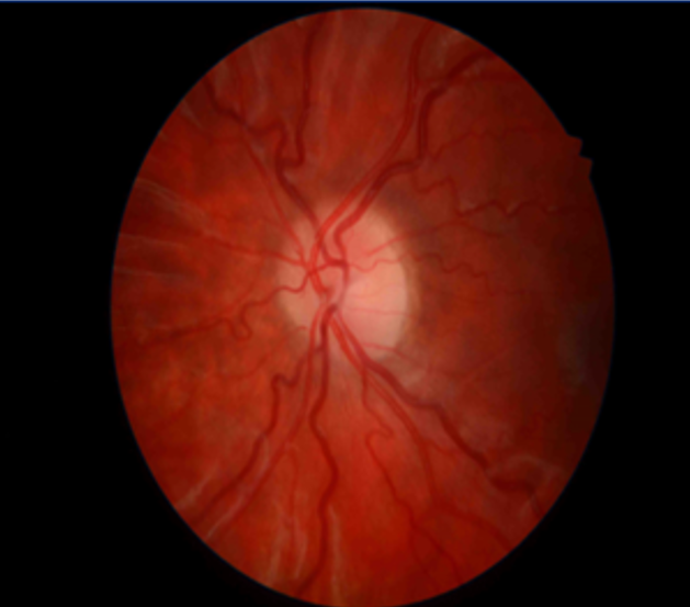
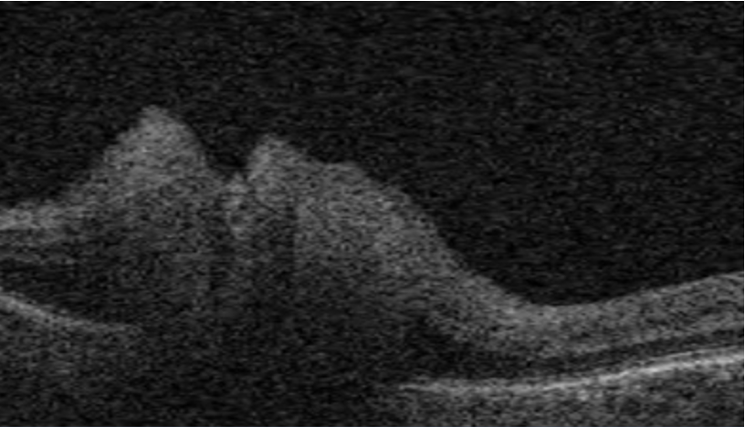
Papilloedema is a term that is exclusively used when disc swelling is secondary to increased intracranial pressure (ICP). (1) . We should distinguish papilloedema from optic disc swelling from other causes, which is named ”optic disc oedema”. As the papilloedema indicates increased intracranial pressure, it is an alarming sign. Potential causes of increased intracranial pressure are brain tumours, central nervous system inflammation (CNS), central venous thrombosis, and idiopathic intracranial hypertension.

It is essential to understand that optic disc oedema is a term used in all other causes when the optic disc swelling is not caused by increased intracranial pressure. For example, due to acute non-ischaemic optic disc neuropathy (NAION), inflammation of the optic disc, infiltration of the optic disc, etc.

02

Symptoms

Patients with papilloedema may have no visual symptoms, especially if the rise in intracranial pressure is acute. The symptoms are mostly related to the increase of the intracranial pressure and manifests with headaches.(4,5) The headaches are worse when the patient wakes up and also get worse when coughing or with another type of Valsalva manoeuvre. In more severe cases, vomiting and nausea can be accompanying symptoms.(4,5) Some patients may experience pulsatile tinnitus, which is hearing rhythmic thumping, whooshing, or throbbing in one or both ears.(6)

Visual acuity in the acute phase is well preserved. Patients who experience visual disturbances complain of visual obscuration (greying-out of their vision, usually in both eyes, significantly when rising from a lying or sitting position) or light flickering. They may also complain of constriction of visual fields and have decreased perception of colour (which is more rare). Furthermore, those with sixth nerve palsy may complain of diplopia.

If you suspect your patient has papilloedema, you should ask specific questions about the type of headache and visual symptoms. Also, ask about recent infections (whether the patient had increased temperature), illnesses, medications, and whether they smoke or not. If your practice has a tonometer, measure the blood pressure to rule out malignant hypertension!

Relative afferent pupillary defect is absent in patients with papilloedema.

03

Clinical signs

Papilloedema can be presented in different stages. It is essential to be familiar with each of them to recognise those cases with early stages of optic disc swelling. You can use the Frisen scale to grade the papilloedema.(7) Be aware that the scale may be subjective but sufficient to lead you to the correct diagnosis.

  • Stage 0 – Normal optic disc
  • Stage 1 – A greyish, C-shaped halo of the optic disc, which preserves the temporal portion of the optic disc. Obscuration of nasal borders of the optic disc. Disruption of the regular radial striation of retinal nerve fibre layers.
  • Stage 2 – The borders of the optic disc are obscured, and the greyish hallo is present in the whole circumference of the optic disc. The optic disc cup is not obscured. The nasal margin of the optic disc is elevated. The central retinal vessels are not obscured.
  • Stage 3 – One or more segments of retinal vessels leaving the disc are obscured. All borders of the optic disc are elevated, which makes the appearance of an increased diameter of the optic disc. The halo is present, which may be irregular and have “finger-like” extensions.
  • Stage 4 – Obscuration of major blood vessels at the disc and all vessels leaving the disc. The optic disc is entirely elevated with the presence of a halo. The cup is not visible.
  • Stage 5 – Obscuration of all vessels at the disc and those leaving the disc. Elevated optic disc with a narrow halo
Image 1. Stage 1 optic disc oedema in a patient with idiopathic intracranial hypertension.
Image 2. The optic disc in a patient with intracranial hypertension three months post-treatment with Diamox tablets.

Bear in mind that papilloedema is always bilateral and generally symmetrical. The cases of idiopathic intracranial hypertension may have the asymmetrical appearance of swelling but still are bilateral. In contrast, optic disc oedema of another origin (e.g., anterior ischemic optic neuropathy) is rarely bilateral.

More severe cases of papilloedema may present with flame-shaped haemorrhages and cotton wool spots. Likewise, patients may develop radial retinal folds, called Paton lines, and choroidal folds.

Image 3. Stage 3 of papilloedema. Temporal to the optic disc edge an intraretinal haemorrhage.

Untreated papilloedema leads to optic disc atrophy, where the optic disc becomes pale without cupping.

If you see optic atrophy in one eye (e.g., due to previous optic disc disease like compressive optic neuropathy) and swelling of the optic disc in the other eye, consider it to be papilloedema. This appearance is called Foster-Kennedy syndrome.(8)

04

Diagnostic procedures

Slit lamp examination/Colour fundus photography – following the Frisen scale, this is a valuable tool to recognize optic disc swelling.

Fluorescein fundus angiogram – a valuable tool to recognise actual leakage from the staining of the optic disc, which is present in the case of optic disc drusen.

Optical coherence tomography – a great tool to recognise subtle swelling of the optic disc. Likewise, monitoring changes, like the beginning of atrophy, is helpful.

Image 4. OCT of optic disc oedema.

Neuro-imaging – MRI and CT scans are essential in papilloedema workup.

05

How to distinguish papilloedema from pseudopapilloedema?

The appearance of optic disc drusen may lead to wrong suspicion of papilloedema, which is an urgent and potentially life-threatening condition. Therefore, it is essential to be familiar with specific clinical signs on distinguishing optic disc drusen-related pseudopapilloedema from the real swelling of the optic disc.


Congestion 

Dilatation 

Retinal vessels 

True optic disc oedema 

Congestion of optic disc vasculature with reddish hue of the disc 

Dilatation of optic disc surface microvasculature/ flame-shapped haemorrhage 

Obscuration of optic disc margin and retinal vessels at the edge of the disc 

Pseudopapilloedema 

No congestion or reddish hue of the disc 

No dilatation of surface microvasculature of the optic disc 

The retinal vessels are clearly visible 

Table 1. Characteristics of papilloedema vs pseudopapilloedema

06

Management and treatment

All patients with papilledema must be urgently referred to an ophthalmologist for further management. It is crucial to treat the cause of the papilledema, which indirectly leads to improved optic disc swelling. Therefore, early recognition of papilloedema is paramount.

07

References

1 Ehlers JP, Shah CP, eds. Papilledema. The Wills Eye Manual: Office and Emergency Room Diagnosis and Treatment of Eye Disease. 5th ed. Baltimore, Md: Lippincott Williams & Wilkins; 2008. 252-254.

2 Dhoot R, Margolin E. Papilledema. InStatPearls [Internet] 2021 Aug 22. StatPearls Publishing.

3 Rigi M, Almarzouqi SJ, Morgan ML, Lee AG. Papilledema: epidemiology, etiology, and clinical management. Eye and brain. 2015;7:47.

4 Sinclair AJ, Burdon MA, Nightingale PG, Matthews TD, Jacks A, Lawden M, Sivaguru A, Gaskin BJ, Rauz S, Clarke CE, Ball AK. Rating papilloedema: an evaluation of the Frisén classification in idiopathic intracranial hypertension. Journal of neurology. 2012 Jul;259(7):1406-12.

5 Scott CJ, Kardon RH, Lee AG, Frisén L, Wall M. Diagnosis and grading of papilledema in patients with raised intracranial pressure using optical coherence tomography vs clinical expert assessment using a clinical staging scale. Archives of Ophthalmology. 2010 Jun 1;128(6):705-11.

6 https://www.pennmedicine.org/for-patients-and-visitors/patient-information/conditions-treated-a-to-z/pulsatile tinnitus#:~:text=What%20Is%20Pulsatile%20Tinnitus%3F,difficult%20to%20concentrate%20or%20sleep.

7 Frisén L. Swelling of the optic nerve head: a staging scheme. Journal of Neurology, Neurosurgery & Psychiatry. 1982 Jan 1;45(1):13-8.

8 https://eyewiki.aao.org/Foster-Kennedy_vs_Pseudo-Foster-Kennedy#:~:text=Foster%2DKennedy%20Syndrome%20is%20characterized,pseudo%2DFoster%2DKennedy%20Syndrome.