Posterior uveitis and panuveitis

Referral priority: urgent

All patients with posterior uveitis or panuveitis must be urgently referred to an ophthalmologist following local guidelines.

Written by
Marko Lukic
Edited by
Svein Tindlund and Jon Gjelle
Published
June 2023

Sections
01
Introduction

Read more
02
Symptoms

Read more
03
Clinical signs

Read more
04
Diagnostic procedures

Read more
05
Management and treatment

Read more
06
References

Read more
01

Introduction

Uveitis is an inflammation of the uveal tract, and is divided into anterior (iris and ciliary body) and posterior components (choroid). The inflammation may affect surrounding tissue like the retina and optic nerve.(1) Uveitis – particularly posterior uveitis – is a common cause of preventable blindness, and is thus deemed a sight-threatening condition. It is responsible for 10% to 15% of all cases of blindness and 30,000 new cases of legal blindness annually in the United States.(2,3) In Western countries, uveitis affects 200 per 100,000 people.(4)4 More than 35% of patients who develop the condition suffer severe visual impairment.(4,5)

There are four types of uveitis, classified by anatomical location:

Type of uveitis  Primary location 
Anterior  Iris, ciliary body, or both 
Intermediate uveitis  Vitreous 
Posterior uveitis  Choroid 
Panuveitis  The entire uveal tract and vitreous 

Then, uveitis is further classified based on the following characteristics:(6)

  • Onset (sudden versus Insidious)
  • Duration (limited, less than 3 months; persistent, more than 3 months)
  • Course (acute, recurrent, or chronic)
  • Laterality (unilateral versus Bilateral)

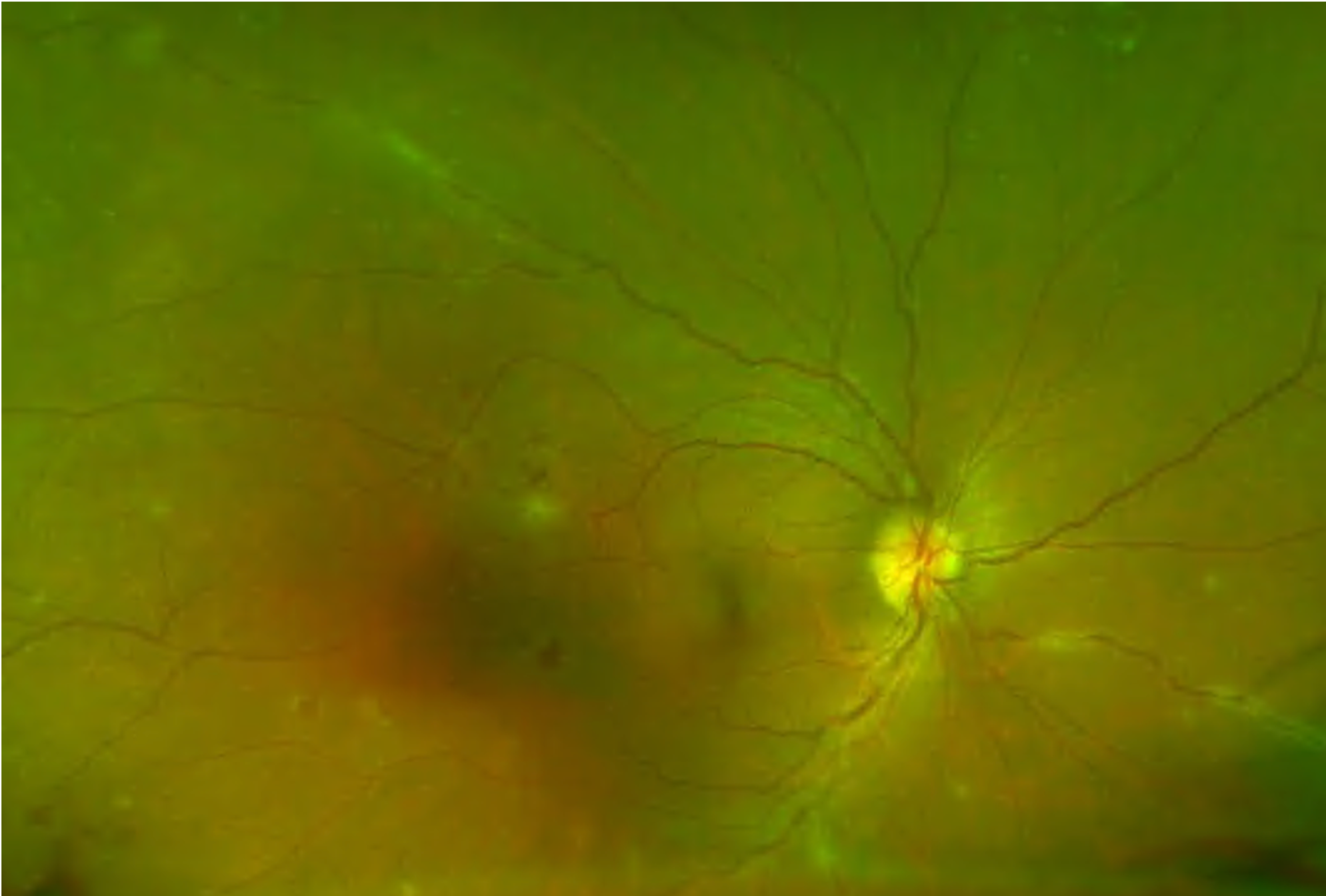
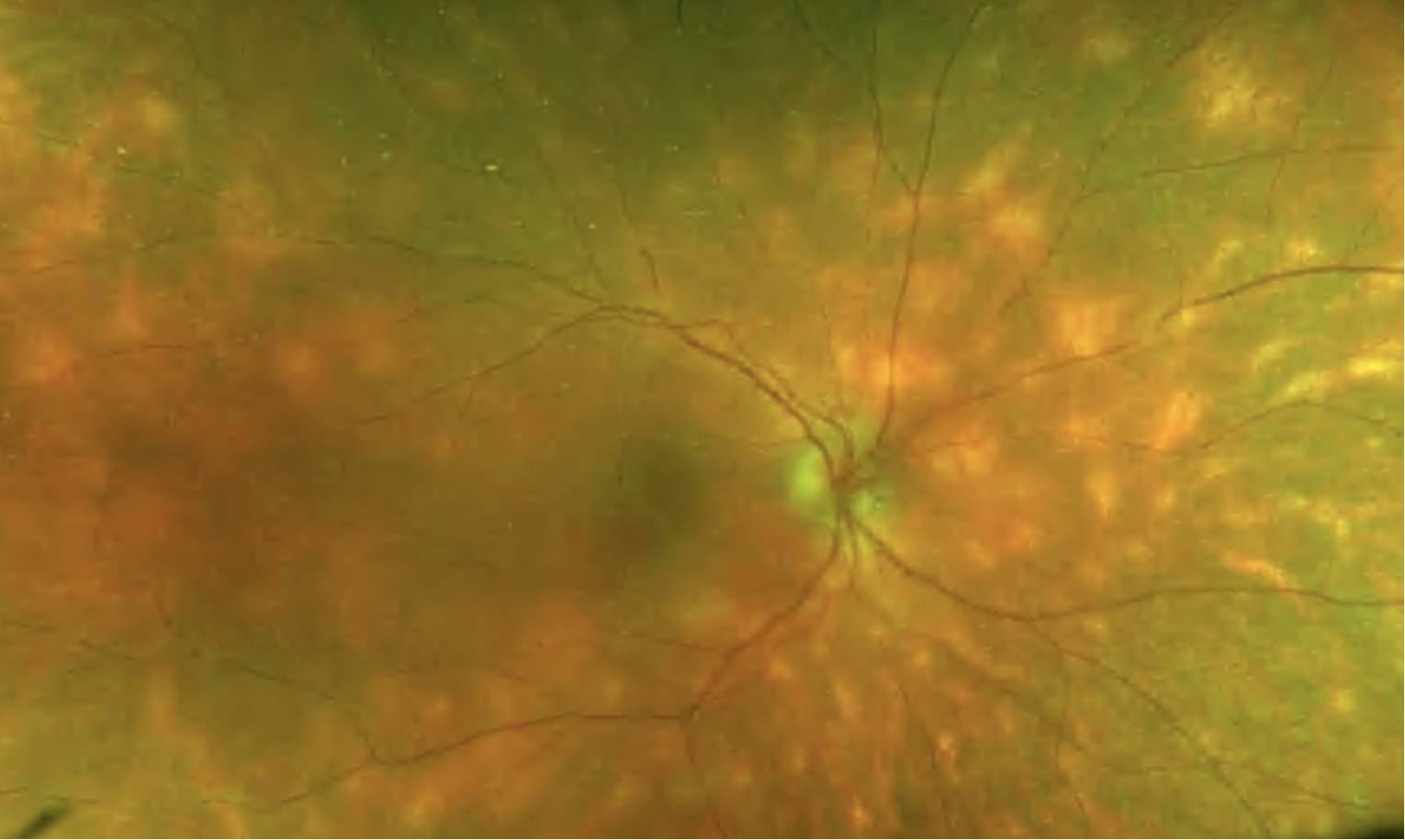
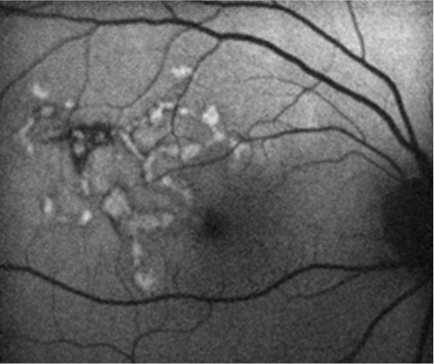
The primary site of inflammation in posterior uveitis is the choroid. It can involve adjacent structures such as the retina, vitreous, optic nerve head, and retinal vessels.(7) Posterior uveitis can be classified based on the cause or the type of lesions. Like in other forms of uveitis, its causes can be infectious (toxoplasmosis, toxocariasis, tuberculosis, syphilis, bartonella, herpes simplex virus, herpes zoster virus, cytomegalovirus, HIV) or non-infectious. Non-infectious types of posterior uveitis may be clinically presented in different ways. Diagnosis is made depending on clinical features and the results of diagnostic testing (i.e., white-dot syndromes, diffuse unilateral subacute neuroretinitis, sarcoidosis, Vogt-Koyanagi-Harada syndrome, sympathetic ophthalmia, serpiginous chorioretinitis, etc.).

The term panuveitis is used when anterior, intermediate, and posterior uveal structures are involved.(8) The causes related to panuveitis are infectious, inflammatory, neoplastic, or traumatic. When neoplasia (retinoblastoma, iris melanoma, and systemic haematological malignancies such as leukaemia and lymphoma) causes panuveitis, it is known under the term “masquerade syndrome”.

02

Symptoms

The symptoms depend on the part of the uveal tract involved. Patients with posterior uveitis complain of blurry vision, floaters, and photopsia. Those with panuveitis may also have eye redness, pain, and photophobia.

It is critical to take a thorough medical history, including present and past personal medical history, surgeries, history of trauma, medicines, and family history. Always ask about arthritis, rashes, shortness of breath, swollen lymph nodes, recent headaches, hearing difficulties, hair loss, pigment changes in the skin, a history of ocular trauma, recent insect bites, sexually transmitted diseases (STDs), tuberculosis exposure, blood in stools, and recent travel.(9-11)

03

Clinical signs

Clinical findings depend mainly on the site of involvement. The clinical signs mentioned in this chapter are based on the site of inflammation.

Anterior portion

Eye redness is present in the form of ciliary injection/flush. However, diffuse redness of conjunctiva may also be present. You should carefully examin the conjunctiva as small nodules may be present.

Keratitic precipitates (KPs) are white blood cells present on the corneal endothelium. Small, white KPs are a sign of the non-granulomatous type of inflammation, while large, yellowish to brown KPs are significant for the granulomatous type of uveitis.

Corneal oedema may be presented and, together with corneal decompensation, may indicate viral aetiology.

Flares and cells in the anterior chamber are signs of intraocular inflammation. Therefore, the cells in the anterior chamber should be graded by severity under high-magnification slit lamp examination in a 1 X 1-mm field of light, as described by The SUN Working Group Grading Scheme for Anterior Chamber Cells, as follows:(8)

  • 0 < 1 cell
  • 0.5 = 1-5 cells
  • 1+ = 6-15 cells
  • 2+ = 16-25 cells
  • 3+ = 26-50 cells
  • 4+ = More than 50 cells

Flare is the presence of proteins in the anterior chamber, and the SUN Working Group proposed the below classification:(8)

  • 0 = None
  • 1+ = Faint
  • 2+ = Moderate (iris and lens details clear)
  • 3+ = Marked (iris and lens details hazy)
  • 4+ = Intense (fibrin or plastic aqueous)


Intermediate portion

Vitreous cells are universal findings in patients with intermediate uveitis. Mostly, they are present in the anterior vitreous. The counting of cells is used, using the same principle as mentioned above for the cells in the anterior chamber.

Vitreous haze may be present in severe cases. The SUN classification is as below (it may be very subjective):(12)

  • Score 0 – No clinical findings
  • Score 1 – Posterior pole clearly visible
  • Score 2 – Posterior pole details hazy
  • Score 3 – Posterior pole very hazy
  • Score 4 – Posterior pole barely visible

Snowballs are aggregates of vitreous cells, mostly located in the inferior vitreous. They are usually white or yellow. They are typical findings in intermediate uveitus, particularly in pars planitis.

Snowbanks are exudates present as plaques in inferior ora serrata. Scleral indentation or indirect ophthalmoscopy is sometimes required to recognise those findings.

Peripheral periphlebitis is inflammation of peripheral venules and may be present in the form of sheathing. It may appear months or years after the initial presentation.

  • Posterior portion Vasculitis is an inflammation of retinal vessels. It may affect veins or arteries.
  • Oedema of the macula and/or optic disc.
  • Retinal haemorrhages.
  • Infiltrates in the choroid (choroiditis) and retina (retinitis) – when combined, the infiltrates are called chorioretinal infiltrates (causing chorioretinitis or retinochoroiditis, depending on what is the primary site of the lesions).
  • Vitreous haemorrhage.
  • Choroidal neovascular membrane.
  • Retinal detachment.

Remember that uveitis may cause late complications like posterior subcapsular cataract, glaucoma, retinal detachment, vitreous haemorrhage, and posterior synechiae.

04

Diagnostic procedures

Slit lamp examination is crucial in the examination of patients with uveitis.

Colour fundus photos are useful in checking posterior eye segments and recognising signs of intermediate and posterior uveitis.

Optical coherence tomography is a golden standard in diagnosing uveitis macular oedema. Uveitis may be complicated with macular oedema, and OCT scans are important in the workup with uveitis patients.

Ultrasound is a very useful tool when optical media is hazy. It is useful in differentiating rhegmatogenous from serous retinal detachment or recognising choroidal thickening.

Fundus fluorescein angiography and indocyanine green angiography (ICG) are invasive angiography modalities and are very useful in diagnosing and monitoring cases of posterior/panuveitis.

Diagnostic vitreous tap procedure – sometimes, taking vitreous sampling and installing treatment has both diagnostic and therapeutic effects.

Image 1. Case of posterior uveitis secondary to sarcoidosis. Note venous sheathing and intraretinal haemorrhages.
Image 2. Case of birdshot choroiditis – note mostly nasal, yellowish, ill-defined deep lesion.
Image 3. Case of posterior uveitis secondary to tuberculosis. The clinical appearance is called serpiginous choroidopathy.
Image 4. Fundus autofluorescence of a case of serpiginous choroidopathy. Note hyperautoflorescense (light grey/white-coloured) lesion, which indicates an active lesion
Image 5. Disturbances of outer retina layers in a case of panuveitis secondary to syphilis.
05

Management and treatment

Advanced laboratory and imaging workups are required in cases of posterior uveitis and panuveitis. It is important to recognise malignant and infectious processes.

Treatment must be done by an experienced uveitis specialist.

Based on the cause, treatment may be specific or non-specific. The specific treatment is against infectious pathogens:

  • Syphilis: Penicillin. Penicillin-sensitive patients can be treated with oral Tetracycline or Erythromycin.
  • Tuberculosis: Standard antitubercular therapy (Isoniazid, Rifampicin, Ethambutol, Pyrazinamide).
  • Toxoplasmosis: Clindamycin, Sulphadiazine, Pyrimethamine, Cotrimoxazole, Atovaquone, Azithromycin – note that treatment depends on the location of the lesion.

Non-specific treatment includes:

  • Cycloplegics
  • Corticosteroids
  • Systemic immunosuppressive agents
    • Alkylating agents – Cyclophosphamide and Chlorambucil
    • Antimetabolites- Azathioprine, Methotrexate, Mycophenolate mofetil
    • T-cell inhibitors – Cyclosporine A, Tacrolimus
  • Biological agents
    • Infliximab, Adalimumab, Etanercept; Inhibit alphaTNF (Tumour Necrosis Factor)
06

References

1 Dunn JP. Uveitis. Prim Care. 2015 Sep. 42 (3):305-23.

2 Nussenblatt RB. The natural history of uveitis. International ophthalmology. 1990 Oct;14(5):303-8.

3 DARRELL RW, WAGENER HP, KURLAND LT. Epidemiology of uveitis: incidence and prevalence in a small urban community. Archives of ophthalmology. 1962 Oct 1;68(4):502-14.

4 London NJ, Rathinam SR, Cunningham ET. The epidemiology of uveitis in developing countries. International Ophthalmology Clinics. 2010 Apr 1;50(2):1-7.

5 Rothova A, Suttorp-van Schulten MS, Frits Treffers W, et al. Causes and frequency of blindness in patients with intraocular inflammatory disease. Br J Ophthalmol. 1996;80:332–336.

6 Rao NA. Uveitis and other intraocular inflammations. Ophthalmology. 2nd ed. Philadelphia: Mosby. 2004:1105-238.

7 Sudharshan S, Ganesh SK, Biswas J. Current approach in the diagnosis and management of posterior uveitis. Indian journal of ophthalmology. 2010 Jan;58(1):29.

8 Standardization of Uveitis Nomenclature (SUN) Working Group. Standardization of uveitis nomenclature for reporting clinical data. Results of the First International Workshop. American journal of ophthalmology. 2005 Sep 1;140(3):509-16.

9 Sanghvi C, Bell C, Woodhead M, Hardy C, Jones N. Presumed tuberculous uveitis: diagnosis, management, and outcome. Eye. 2011 Apr;25(4):475-80.

10 Kamal S, Kumar R, Kumar S, Goel R. Bilateral interstitial keratitis and granulomatous uveitis of tubercular origin. Eye & Contact Lens. 2014 Mar 1;40(2):e13-5.

11 Abderrahim K, Chebil A, Falfoul Y, Bouladi M, Matri LE. Granulomatous uveitis and reactive arthritis as manifestations of post-streptococcal syndrome. International ophthalmology. 2015 Oct;35(5):641-3.

12 Davis JL, Madow B, Cornett J, Stratton R, Hess D, Porciatti V, Feuer WJ. Scale for photographic grading of vitreous haze in uveitis. American journal of ophthalmology. 2010 Nov 1;150(5):637-41.