Posterior vitreous detachment

Referral priority: Low or urgent – depending on symptoms and clinical signs

Closely follow local guidelines for dealing with posterior vitreous detachments and retinal detachments. Patients with signs of vitreous haemorrhage, pigment in the vitreous, retinal tears or retinal detachments must be urgently referred to an ophthalmologist.

Written by
Marko Lukic
Edited by
Svein Tindlund and Jon Gjelle
Published
June 2023

Sections
01
Introduction

Read more
02
Symptoms

Read more
03
Clinical signs

Read more
04
Diagnostic procedures

Read more
05
Management and Treatment

Read more
06
References

Read more
01

Introduction

The vitreous is a clear gel that fills the space between the lens, ciliary body anteriorly, and retina posteriorly.(1) It comprises approximately 80% of the volume of the eye, and consists of approximately 98% water and 2% proteins, and an extracellular matrix. Collagen is the major structural protein (type II 75% and type IX 15%).(1,2)

The strongest attachment points of the vitreous are at the optic nerve, macula, ora serrata, and major retinal blood vessels.(3) The equatorial and posterior vitreous interface consists of the posterior vitreous cortex, internal limiting membrane (ILM), and the intervening extracellular matrix. The intervening extracellular matrix is a macromolecular complex that glues the ILM and posterior cortex. It is composed of fibronectin, laminin, chondroitin sulfate, and other structures of the extracellular matrix.(1)

Posterior vitreous detachment (PVD) is a separation between the neurosensory retina and the posterior vitreous cortex.(4) The initial event is liquefaction and syneresis of the central vitreous (it becomes liquid and shrinks due to age).(1,2) A rupture develops in the posterior hyaloid (or vitreous cortex) through which liquefied vitreous flows into the retrovitreous space, separating the posterior hyaloid from the retina.

The prevalence of PVD increases with age and the eye’s axial length. The general onset of the event is between the ages of 60-80, with no difference between men and women. Risk factors for developing PVD earlier in life are high myopia, blunt ocular trauma, inflammation of the eye, and previous eye surgery, particularly cataract surgery.
02

Symptoms

The posterior vitreous detachments start as a partial event, where they first begin to separate from the perifoveal area. At that stage, the patients are asymptomatic or have mild symptoms.

Presenting symptoms include entoptic phenomena such as floaters, changes in the pattern of floaters, and photopsia (flashing lights).

Floaters are the most common complaint and result from vitreous opacities such as blood, glial cells, or aggregated collagen fibres torn from the margin of the optic disc. They move with vitreous displacement during eye movement and scatter incident light, which casts a shadow on the retina that is perceived as a grey structure resembling “hairs”, “flies”, or ”spiderwebs”. Floaters may continue indefinitely, although they usually gradually diminish over time.

During the first few weeks of PVD, the floaters come with flashing lights which are most noticeable in dim light. The flashing lights are vertical and present in temporal fields of the vision. They are secondary to physical stimulation of the retina by vitreoretinal traction. Hence, it clearly explains why they should be taken seriously, and why they are warning signs of potential retinal tear formation.

Be aware that mild floaters may be normal findings. However, sudden appearance of floaters (sometimes patients say these are new floaters if they have had mild ones before), especially in combination with flashing lights, are signs of PVD.

03

Clinical signs

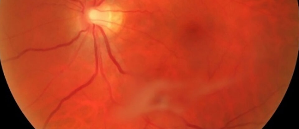
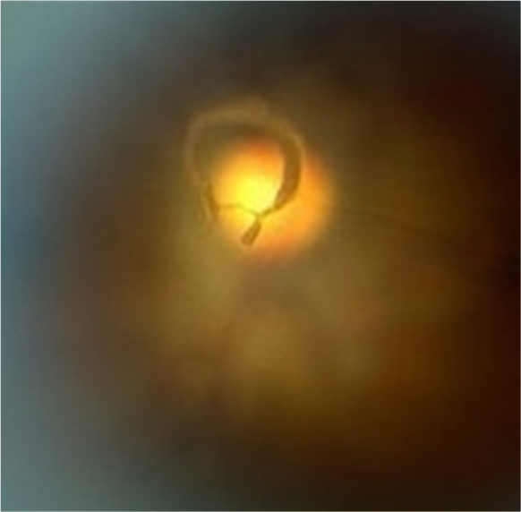
The posterior vitreous detachment is usually diagnosed with a dilated eye examination. The Weiss ring, a concentric ring at the projection of the optic disc, is a sign of a complete PVD.

The presence of vitreous haemorrhage and pigment cells in the vitreous are warning signs of retinal tear and/or retinal detachment. You should refer those patients urgently referred to an ophthalmologist, even if you cannot find the retinal tear on eye examination or if you consider the retina is fully attached.

Be aware that 15% of symptomatic PVD patients develop a retinal tear. If there is the presence of vitreous haemorrhage, the likelihood of the presence of a retinal tear is between 50-70%.(5)

04

Diagnostic procedures

Slit lamp examination – the most common way of identifying PVD and checking the retina for retinal tears and/or retinal detachment.

Image 1. The Weiss ring is a sign of complete PVD (detachment of the posterior vitreous from the optic disc). It is seen in the vitreous cavity at the projection of the optic disc.

Ultrasound – a great tool to distinguish PVD from retinal detachment (RD). It is very useful when there is dense vitreous haemorrhage to distinguish the PVD from RD.

Image 2. An ultrasound image of PVD. Notice a thin line detached from the retina. The line is also detached from the optic nerve, which is one of the distinguishing signs of retinal detachment.

Optical coherence tomography – if the vitreous is clear and you cannot see the PVD on a slit lamp, this imaging modality nicely shows the separation of the hyaloid membrane of the neurosensory retina.

Image 3. An OCT image of the detached posterior hyaloid membrane from the ILM.
05

Management and Treatment

If the patient is symptomatic (new onset of floaters and flashing lights), and if there is no presence of retinal tear and retinal detachment, you should review the patient in 2 weeks – or earlier if the symptoms progress (new floaters, increased intensity of flashing lights, changes in visual fields like new black/grey shadow). You should review patients with mild PVD in a month, and explain the warning signs.

There is no treatment for PVD. However, the complications of PVD, like vitreous haemorrhage, retinal tear, rhegmatogenous retinal detachment, and tractional and exudative retinal detachment require urgent treatment.

06

References

1 S.J. Ryan, C.A. Puliafito, J.L. Davis, J.M. Parel, P. Milne Retina (4th ed.), Elsevier Mosby, Philadelphia, PA (2006)

2 De Smet MD, Elkareem AM, Zwinderman AH. The vitreous, the retinal interface in ocular health and disease. Ophthalmologica. 2013;230(4):165-78.

3 Tsui I, Pan CK, Rahimy E, Schwartz SD. Ocriplasmin for vitreoretinal diseases. Journal of Biomedicine and Biotechnology. 2012 Jan 1;2012.

4 Duker JS, Kaiser PK, Binder S, de Smet MD, Gaudric A, Reichel E, Sadda SR, Sebag J, Spaide RF, Stalmans P. The International Vitreomacular Traction Study Group classification of vitreomacular adhesion, traction, and macular hole. Ophthalmology. 2013 Dec 1;120(12):2611-9.

5 D’Amico DJ. Primary retinal detachment. New England Journal of Medicine. 2008 Nov 27;359(22):2346-54.