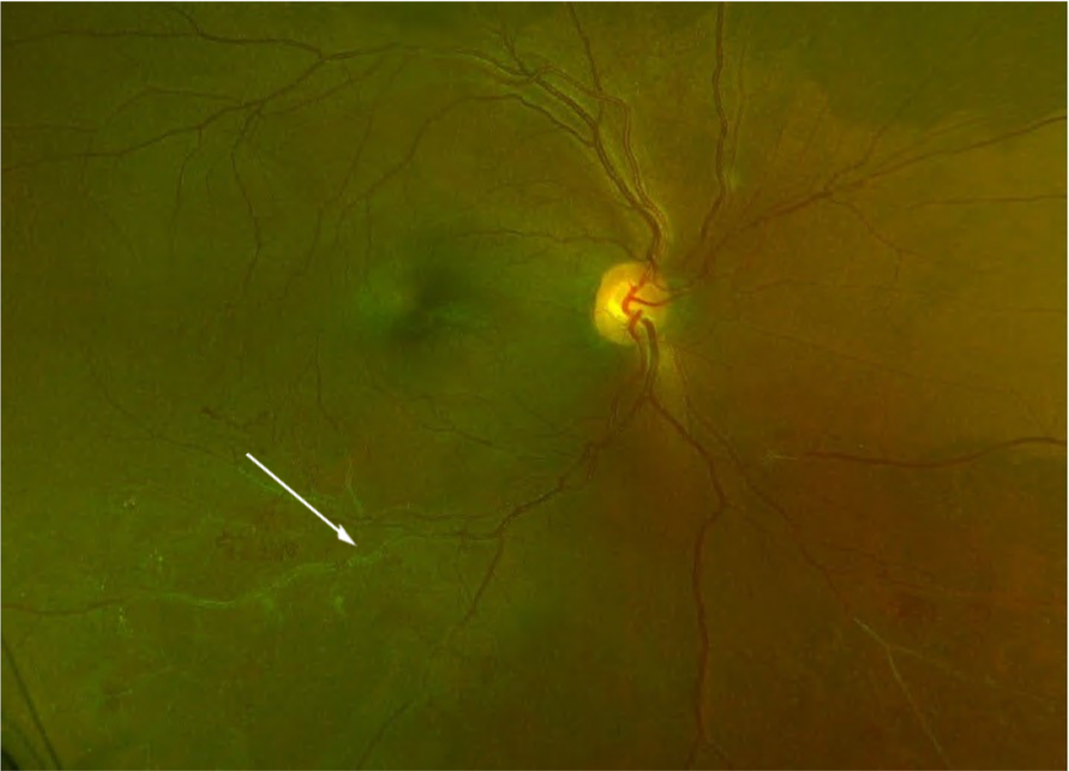
Retinal vein occlusion (RVO) is a common retina vascular disorder and the second most common cause of blindness from retinal vascular disease after diabetic retinopathy.(1) It is classified based on the location of the occlusion. The cause of the occlusion differs, and in central retinal vein occlusion (CRVO), the cause is a thrombus which occludes the central retinal vein near the lamina cribrosa.(2) Hemi-retinal vein occlusion (HRVO) is a form of CVRO where the inferior or superior branch of the retinal vein is occluded, and half of the retina affected.
The branch retinal vein occlusion (BRVO) occurs when a thrombus occurs at the arteriovenous crossing point, secondary to atherosclerosis of the retinal artery causing compression of the retinal vein.(3) There are two major subtypes of BRVO: major and macular. The incidence of BRVO is most common in the superior temporal quadrant (58.1-66%), followed by the inferior temporal quadrant (29%), and least common in the nasal quadrants (12.9%).(4,5) Macular BRVO involves the superior macular region in 81% of cases and the inferior macular region in 19% of cases.(6)
The aetiology and risk factors of CRVO and BRVO differ.According to the Eye Case-Control Study Group, the risk factors for CRVO are hypertension, open-angle glaucoma, and diabetes mellitus, while the risk factors for BRVO are hypertension, high body-mass index, open-angle glaucoma, and cardiovascular disease.(7,8)
Further studies proved that other conditions are risk factors for developing central retinal vein occlusion:(8-11)
- carotid insufficiency
- hematologic alterations (hyperviscosity syndrome, multiple myeloma, blood dyscrasias (polycythaemia vera, lymphoma, leukaemia, sickle-cell disease or trait), anaemia, elevated plasma homocysteine, factor XII deficiency, antiphospholipid antibody syndrome, activated protein-C resistance, protein-C deficiency, protein-S deficiency)
- systemic lupus
- HIV
- syphilis
- some medications (oral contraceptives, diuretics)
- dehydration
- pregnancy
There is conflicting evidence that hypercoagulable conditions are risk factors for BRVO, while vasculitis is indeed one of the risk factors.(12)
Most patients with CRVO are older than 65 years. Most cases are unilateral, with approximately 6%-14% of the cases bilateral. BRVO is three times more common than CRVO. Men and women are affected equally, mostly between the ages of 60 and 70.(13) A large study from 2010 reported the prevalence of BRVO to be 2.8 per 1,000 in whites, 3.5 in blacks, 5.0 in Asians, and 6.0 in Hispanics, and the prevalence of CRVO to be 0.88 per 1,000 in whites, 0.37 in blacks, 0.74 in Asians, and 1.0 in Hispanics.(14)
The pathogenesis of CRVO is believed to follow the principles of Virchow’s triad for thrombogenesis, involving vessel damage, stasis, and hypercoagulability.(14) The central retinal vein and artery share a common adventitial sheath at arteriovenous crossings, posterior to the lamina cribrosa, so that atherosclerotic changes of the artery may compress the vein and cause CRVO.(15)
The association between BRVO and arteriovenous crossings has been proved in multiple studies. In almost all cases of BRVO (97.6-100%) the thick-walled artery is found anterior to the thin-walled vein.(16-18) The artery and vein also share a common adventitial sheath at these crossings, contributing to the predisposition of vein occlusion at these crossings. Arteriolar sclerosis increases the rigidity of the artery and further supports the mechanical basis of BRVO at arteriovenous crossings.(16,19) Mechanical compression of the vein by the rigid artery results in turbulent blood flow at arteriovenous crossings, resulting in venous intima-media and endothelial damage, which leads to vein occlusion.(5,20)
In addition, both BRVO and CRVO can be divided according to perfusion status, and classified as non-ischaemic and ischaemic. It is essential to be aware of the signs and symptoms of ischaemic form as it risks new vessel development and has a poorer visual prognosis.(21)


