Rhegmatogenous retinal detachment

Referral priority: Urgent

All patients with a retinal tear or retinal detachment must be urgently referred to an ophthalmologist or hospital emergency room following local guidelines.

Written by
Marko Lukic
Edited by
Svein Tindlund and Jon Gjelle
Published
June 2023

Sections
01
Introduction

Read more
02
Symptoms

Read more
03
Clinical signs

Read more
04
Diagnostic procedures

Read more
05
Management and Treatments

Read more
06
References

Read more
01

Introduction

Retinal detachment is the separation of the neurosensory retina from the retinal pigment epithelium (RPE). It is a sight-threatening condition with an incidence of 1 in 10,000.(1,2) Furthermore, retinal detachment is more common in men, and mainly present in people between 40 and 70 years old.

Retinal detachment occurs when fluid accumulates in the virtual subretinal space. The classical categorisation of retinal detachment is rhegmatogenous, tractional, and exudative (serous). The rhegmatogenous retinal detachment (RRD) is the most common, and it occurs when there is a full-thickness retinal break secondary to vitreoretinal traction (most often during the process of posterior vitreous detachment (PVD)). The break allows fluid to enter the subretinal space and separates the neurosensory retina from the RPE.

Three elements need to be present for the rhegmatogenous retinal detachment:

  1.  Abnormal mobility of partially liquefied vitreous.
  2. Tractional elements that cause retinal tear formation.
  3. Retinal break.

These are the risk factors for rhegmatogenous retinal detachment: lattice degeneration, peripheral retinal breaks, pathologic myopia, trauma, previous intraocular surgery, aphakia, family history, and previous retinal detachment. The peripheral retinal degenerations are important risk factors where lattice degeneration takes the primary position.(3)
02

Symptoms

Patients report symptoms of the PVD, like new-onset floaters and photopsia. Those symptoms may implicate the presence of a retinal tear, which is the cause of retinal detachment. Patients who have developed retinal detachment may notice a greyish shadow (black curtain) in the visual fields. The vision could decrease significantly if the retinal detachment affects the macula or fovea.

If you suspect your patient has retinal detachment, check whether they have one of the risk factors.

Be aware that patients who develop new-onset floaters and photopsia have a 15% risk of developing retinal tears.

03

Clinical signs

It is crucial to be familiar with the term retinal break and how to recognise one. The retinal break is referred to as two clinical manifestations: a retinal tear and a retinal hole. The retinal tears are related to vitreoretinal traction posterior to the vitreous base and occur during the PVD. Retinal holes are the result of localised retinal atrophy and often have the presence of an operculum. The operculum is a sign of vitreoretinal relief in that area.(4-6)

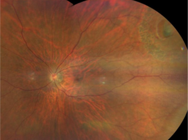
Image 1. Superior temporal retinal tear with operculum in the vitreous. No retinal detachment can be seen.
Image 2. Superior temporal U-shaped retinal tear. Pigmented laser scars can be seen surrounding the tear. No retinal detachment can be seen.

The most common retinal tears are so-called “horse shoes” or “U-shaped” ones. They are commonly present at the posterior border of the vitreous base, although they can be present near the equator. The most common location is the superior temporal quadrant (60%), following the superior nasal quadrant. Of all RRD cases, 50% have more than one retinal break.(7) The actual tear consists of two anterior extensions called horns, which run forward from the apex of the flap. The flap represents torn retinal tissue where during the tractional moment, the retina is pulled anteriorly and torn. The base of the flap remains attached.

The warning signs of retinal tears are vitreous haemorrhage and pigment in the anterior vitreous. The pigment seen in the anterior vitreous is seen as pigmented granules and originates from the RPE. This is called Shaffer’s sign or tobacco dust – and when this is present, the likelihood of a retinal tear is 90%.

There are four Lincoff rules to recognise the location of the retinal break. They could be helpful in everyday clinical practice.(8)

  • Rule 1: When the detachment is in the superior temporal or nasal quadrants, the break is within 1,5 clock hours of the highest border of the detachment (in 98% of cases).
  • Rule 2: For total or superior detachments that cross the 12 o’clock meridian, the retinal break is at 12 o’clock or 1,5 hours within (93% of cases).
  • Rule 3: For inferior retinal detachments, the higher side of the detachment indicates the side of the break (95% of cases).
  • Rule 4: When there is the presence of bullous and symmetrical inferior detachment, there is a small retinal break at 12 o’clock.

The characteristic appearance of rhegmatogenous retinal ’detachment differentiates it from a tractional or exudative detachment. A rhegmatogenous retinal detachment has a corrugated appearance and undulates with eye movements. The main characteristic is, of course, the presence of a retinal break. Be aware that retinal breaks may be obscured by haemorrhage or vitreous opacities or can be too small to be visualised.

The retinal detachment is classified based on the macular involvement. If the macula or fovea is involved, it is called macula off-retinal detachment. If the macula is not involved, which means that the macula is still attached, it is called the macula on-retinal detachment.

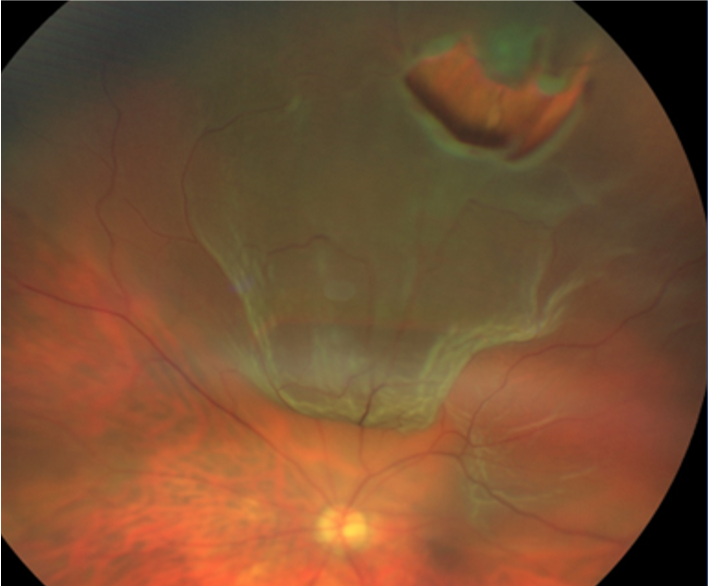
Image 3. Rhegmatogenous retinal detachments with macula on. Notice the corrugated appearance of the detached retina. A tear in the superior temporal quadrant is seen.
04

Diagnostic procedures

Slit lamp examination with auxillary lenses – in the case of clear optic media, the rhegmatogenous retinal detachment should be easily recognised. It is important to be familiar with the pathophysiology of the condition. Retinal detachments can be visible on red reflex testing as a grey shadow and will also be visible when examining the fundus with auxiliary lenses (such as 90D).

Ultrasound – very useful when media opacities are not clear (i.e., dense vitreous haemorrhage). It differentiates posterior vitreous detachment (posterior hyaloid membrane) from the detached retina. Sometimes, a retinal tear can be recognised with an ultrasound.

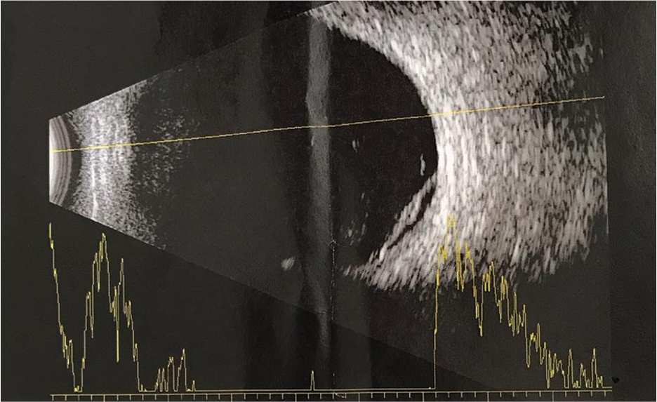
Image 4. Ultrasound image of the detached retina. Notice a hyperreflective, thick layer detached from the choroidal layer.

Optical coherence tomography – useful to differentiate retinal detachment from retinoschisis. It is more useful when the changes are located more centrally. However, a good technician should be able to catch the more peripheral lesion, which may then help recognise retinal detachment. Likewise, OCT could be useful to differentiate rhegmatogenous retinal detachment from two other subtypes: tractional and exudative.

Image 5. OCT scan of a detached retina. Notice that the whole neurosensory retina is detached from the RPE, a distinctive sign from the retinoschisis.
05

Management and Treatments

Generally, all patients with a retinal break (tear or hole) should be reviewed by an ophthalmologist. The decision to treat a retinal tear is based on several factors, including symptomatology, age and systemic health, refractive error, location, type, size of the break, status of the fellow eye, lens status, and family history of retinal breaks or retinal detachments.

Symptomatic retinal tears should be treated immediately, and the treatment of choice is laser retinopexy. The laser treatment aims to create chorioretinal adhesions and prevent the spreading of subretinal fluid.

The treatment of asymptomatic retinal tears is controversial, and it is out of the scope of this chapter. Generally, retinal tears which are demarcated with pigment may be considered chronic and can only be monitored.

Retinal detachment requires surgical treatment. Three procedures may be used: pneumatic retinopexy, scleral buckling, or vitrectomy. Nowadays, vitrectomy is the most used procedure among vitreoretinal surgeons.

Macula on-retinal detachment is urgent, and the patients should undergo surgery within 24 hours. It is acceptable that macula off-retinal detachment surgery is delayed for a couple of days. However, there is evidence that the duration of macula detachment affects the final visual acuity outcomes.(9)

06

References

1 Haimann MH, Burton TC, Brown CK. Epidemiology of retinal detachment. Archives of ophthalmology. 1982 Feb 1;100(2):289-92.

2 Go SL, Hoyng CB, Klaver CC. Genetic risk of rhegmatogenous retinal detachment: a familial aggregation study. Archives of ophthalmology. 2005 Sep 1;123(9):1237-41.

3 Gupta OP, Benson WE. The risk of fellow eyes in patients with rhegmatogenous retinal detachment. Current opinion in ophthalmology. 2005 Jun 1;16(3):175-8.

4 Byer NE. What happens to untreated asymptomatic retinal breaks, and are they affected by posterior vitreous detachment?. Ophthalmology. 1998 Jun 1;105(6):1045-50.

5 Byer NE. Clinical study of retinal breaks. Transactions-American Academy of Ophthalmology and Otolaryngology. American Academy of Ophthalmology and Otolaryngology. 1967;71(3):461-73.

6 Byer NE. The natural history of asymptomatic retinal breaks. Ophthalmology. 1982 Sep 1;89(9):1033-9.

7 https://emedicine.medscape.com/article/1224737-clinical#b4

8 Diagne JP, De Medeiros ME, Ka AM, Samra A, Diallo HM, Sy EH, Aw A, Gueye A, Sow S, Diop LA, Ndiaye JM. Rhegmatogenous retinal detachment: Topography of breaks and agreement with ’lincoff’s rules. Journal Francais D’ophtalmologie. 2019 Nov 21;43(1):31-4.

9 Lee CS, Shaver K, Yun SH, Kim D, Wen S, Ghorayeb G. Comparison of the visual outcome between macula-on and macula-off rhegmatogenous retinal detachment based on the duration of macular detachment. BMJ Open Ophthalmology. 2021 Mar 1;6(1):e000615.