Apr 19, 2021 • 30 minutes read

How to get started with myopia management

Introcution

Written by
Pelsin Demir

Myopia is becoming more common in children across the world, and the onset of myopia is earlier than ever, with more children progressing to high myopia. We also associate high myopia with higher future risks to vision. There are estimates that by 2050, myopia will affect almost half of the world’s population! If we look at high myopia, we know from various research studies that it’s increasing globally. By 2050, at least 1 billion people will be highly myopic, which means that at least 1 billion people will be at risk of permanent vision impairment in the future.

In this handbook, you’ll learn all about myopia: the basics, classification, prevalence, differences across regions of the world, potential consequences of high myopia and the associated ocular complications for the patient, practitioner, and community as a whole. Why do people develop myopia? What are the risk factors for developing myopia? How can we slow down the progression of myopia? These are some of the questions that we’ll dive into. The myopia area is complex and sometimes complicated. With this handbook, you’ll better know how to deal with your patients with myopia.


Chapters

Five things you should know about myopia management

01
Speaking of myopia…

How to classify myopia

Go to chapter
02
Higher isn’t always better

What are the risks and dangers of high myopia?

Go to chapter
03
Will half the world be myopic by 2050?

Get to know the risk factors for developing myopia

Go to chapter
04
Take care of your little patients

How to build a risk profile

Go to chapter
05
Get to know your options for treatment

How to manage myopia

Go to chapter
01

Speaking of myopia…

How to classify myopia

Myopia is commonly known as the eye’s refractive state, in which light comes to focus in front of the retina in the absence of accommodation. This condition results in blurred vision, mainly at far distances. There are two types of myopia. If the eye’s axial length is too long for its optical power (cornea and crystalline lens), it is called axial myopia. On the other hand, if the optical power is too strong for its axial length, it is called refractive myopia. The most common type is axial myopia. There are rare cases where the type of myopia is due to refractive myopia.

We can define and classify myopia in several ways, and that is where it gets complicated. You can easily get confused by all the different terms. In this chapter, I’ll go through every single classification: by degree, age of onset or clinical entity.

Schematic representation of refractive and axial myopia. Axial myopia is the most common type, where the axial length is too long compared to the optical power of the eye (cornea and crystalline lens).

Classification by degree

Classifying myopia by degree is the most common way of classification, and most likely, the one you use at your practice daily. The consideration of myopia sometimes differs when reading studies or literature. For instance, some studies consider a patient as myopic when their refractive error is -0.50 D or more, but others may define their patient as myopic when their refractive error is -0.75 D or more. The difference may be due to the age of the patient or the methodology of the examination. If the patient is a child and undergoes cycloplegic refraction, studies usually define myopia as -0.50 D or more. However, if the patient is a child but doesn’t undergo cycloplegic refraction, studies usually define myopia as -0.75 D or more. The reason some studies increase the definition of myopia is to eliminate artificial myopes with uncontrolled accommodation.

At this point, according to the World Health Organization (WHO), the criteria for myopia is -0.50 D or worse. This means that as soon as a patient has -0.50 D or worse, they’re considered myopic and classified in the ‘low myopia’ category. Subsequently, if the refractive error is between -2.00 D to -5.00 D, it’s classified as ‘mild myopia’, and if it’s -5.00 D or worse, it’s classified as ‘high myopia’. In earlier days, high myopia has been classified as myopia worse than -5.00, -6.00, or even -8.00 D. The reason that -5.00 D is now considered as high myopia is because the risk of complications increases significantly at this level. Also, an uncorrected myope with a refractive error of -5.00 D meets the WHO blindness criteria. Therefore, I would recommend these categories if you want to classify myopia by degree.

  • Low myopia: -0,50 D to −2,00 D
  • Mild myopia: -2,00 D to -5,00 D
  • High myopia: −5.00 D or more

Classification by age of onset

Another way of classifying myopia is by age of onset. Here we can distinguish four categories:

  • Congenital myopia (present at birth and persisting through infancy)
  • Youth-onset myopia (< 20 years of age)
  • Early adult-onset myopia (20-40 years of age)
  • Late adult-onset myopia (>40 years of age)

Classification by clinical entity

Last but not least, we have classification of myopia by clinical entity. I’ll describe them one by one.

Congenital myopia
Congenital myopia is present since birth, and the patient is usually diagnosed at 2-3 years of age. We see this more frequently in children who were born prematurely or with various birth defects. The refractive error is about -8.00 D to -10.00 D and remains constant. Congenital myopia can sometimes be associated with conditions such as cataract, microphthalmos, aniridia, and megalocornea. If it’s is unilateral, the patient may develop amblyopia, which can lead to strabismus. If congenital myopia is bilateral, the patient may have difficulties with distant vision and holding objects very close. It’s best if congenital myopia is corrected early, to help children develop normal distance vision and perception of the world. The child will undergo retinoscopy under full cycloplegia and prescribed with full correction.

Simple myopia
Simple myopia, also named physiological or school myopia, is due to a physiological error and isn’t associated with any eye diseases. The clinical picture of simple myopia is rarely present at birth, and the patient is born hyperopic and becomes myopic at the age of 7 to 10 years. The progression of myopia stabilises around the mid-teens, and the patient usually reaches a refractive error of around -5.00 D and never exceeds -8.00 D. The reason for developing simple myopia is often because of spending many hours looking at close distance and spending too little time outdoors.

Late-onset myopia
Patients with late-onset myopia are typically adults of 20 to 40 years of age, mostly students or workers who spend a lot of time in front of screens without taking any breaks. Their myopia develops slowly. A combination of accommodative anomalies, many near activities, and too little daylight may cause late-onset myopia.

Nocturnal myopia
Nocturnal myopia or night myopia occurs only under conditions of dim illumination or darkness and primarily appears due to a combination of increased accommodative response and low levels of light. In the dark, both insufficient contrast and increased spherical aberrations result in an inadequate accommodative stimulus. So, the eyes don’t focus on infinity but assume the intermediate dark focus for an accommodative position. The main symptom of nocturnal myopia is blurred distance vision in dim illumination. Patients may complain of difficulty seeing road signs when driving at night.

Pseudo-myopia
Pseudo-myopia, also called artificial myopia, occurs due to an accommodative disorder. The patient has an excessive accommodation and spasm of accommodation. Be careful with pseudo-myopic patients! They can really fool you, and you need to be attentive. Pseudo-myopia is generally encountered in younger emmetropes or slightly hyperopes who spend much time doing near work or activities. The patient complains about blurry vision at far distances that’s transient but greater, especially after near activities. This is what happens: when the patient is viewing near for a longer time, without taking any breaks, it causes hypertonicity of the ciliary body of the eye. Therefore, an emmetropic or slightly hyperopic patient may clinically appear to be myopic. If the patient is myopic, it may still seem to be more than it already is.

So, now you may wonder: what do I do with a pseudo-myopic patient? Or how can I identify them? Well, in most of the cases, the autorefractor gives you a high negative refractive error value such as -6.00 D or -7.00 D, and you think: “oh, this is a high myope”. You take the patient’s history, and he or she says: “This is the first time I will undergo an eye examination. I just started a new job with a lot of screen time, or I study all day, and I can’t see at far distances at the end of the day. It gets clear after a while, but this repeats itself every day”. Then you measure their visual acuity, and they see 20/20, which doesn’t make any sense, right? These are all red flags! What you have to do now is relaxing the patient’s accommodation, either with cycloplegia or letting the patient wear trial frames with at least +2.00 D for approximately 40 minutes. After that, you perform a refraction and always prescribe plus lenses.

Induced myopia
The natural history of induced myopia depends on the initiating condition or agent. A refractive shift towards myopia after the age of 60 is usually associated with the development of nuclear sclerosis of the crystalline lens – therefore, it’s a form of induced myopia. Induced myopia is often temporary and reversible. Variability in blood sugar levels may also cause induced myopia. It’s essential that you ask questions about the patient’s blood sugar level and whether this is stable. High blood sugar levels may show a more myopic refractive error, and low blood sugar levels may give a more hyperopic refractive error. Patients with induced myopia also report blurred distance vision. The time course of the distance blur depends on the agent or condition that has induced myopia. Whether other symptoms are present depends on the cause of the induced myopia.

Degenerative myopia
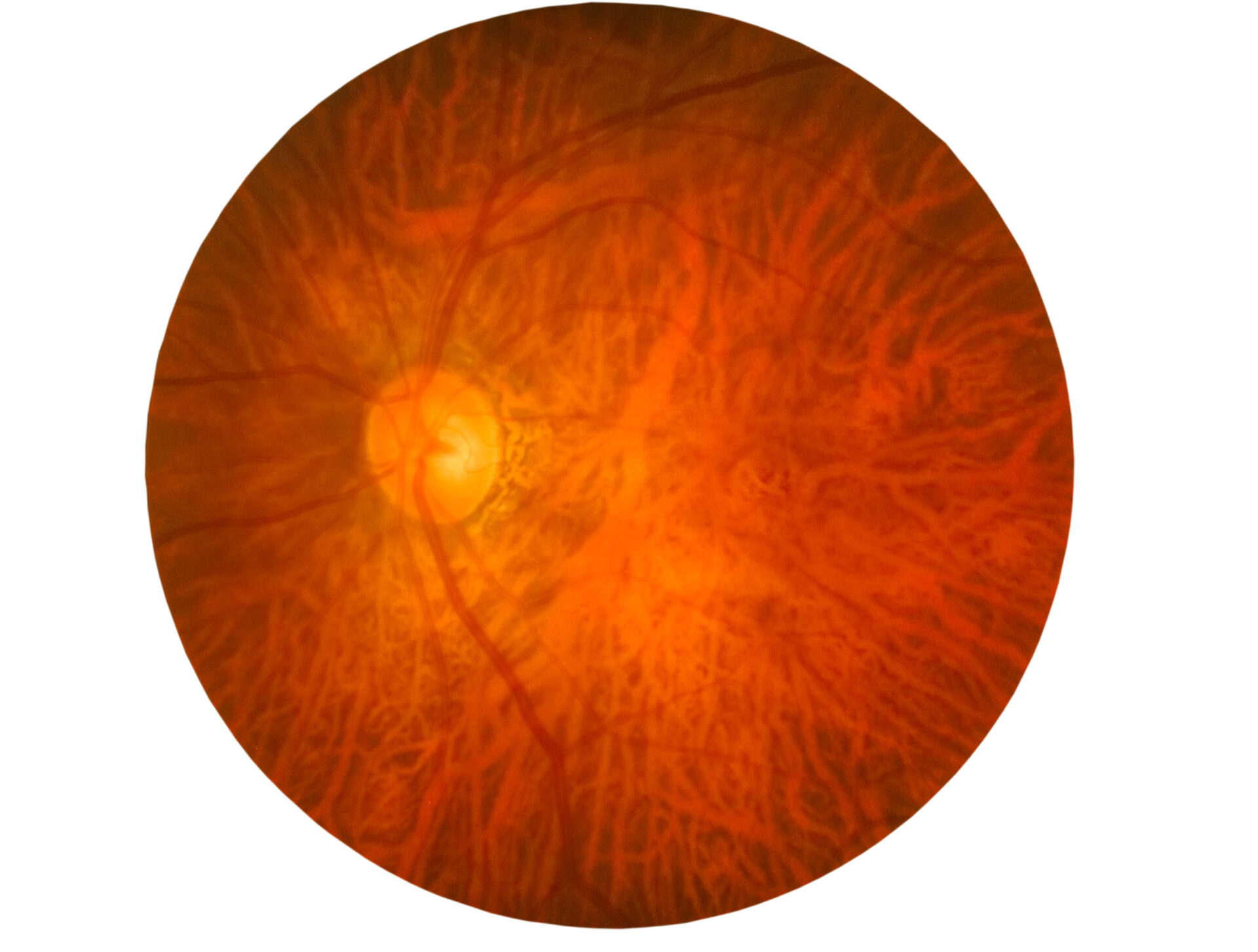
Degenerative myopia appears because of the rapid growth of the axial length, which grows outside the normal biological variations of development. This rapid development may also be associated with pathological changes in the eye. Elongation of the eye causes stretching of the retina and the tissue behind the retina. We see this as a tessellated appearance in fundus pictures and increased visibility of the choroidal vasculature. The peripheral retina gets affected as well and produces characteristic changes of degenerative myopia.

If a patient has degenerative myopia, they have to hold near-point objects quite close to the eyes due to the magnitude of uncorrected myopia. The patient may notice flashes of light or floaters associated with vitreoretinal changes. If pathological posterior segment changes have affected the retinal function, the patient may have a history of vision loss and, perhaps, uses low-vision services or devices. The refractive error increases by as much as 4.00 D yearly and stabilises at about 20 years of age. Occasionally, it may progress until the mid-30s and frequently result in myopia of -10.00 D to -20.00 D. Patients with degenerative myopia may also express concern about the high power of their optical correction. They often have some discomfort due to the weight or inconvenience of the correction. Severe congenital myopia during infancy or high myopia typically becomes degenerative myopia.

Remember that there’s no golden standard of classification, and it depends on who you’re talking to. It’s important to be aware of the differences to avoid causing any miscommunication between your colleagues or other professionals.

Conclusion

As there are so many ways to classify myopia, you can easily get confused and cause miscommunication when talking to fellow optometrists or ophthalmologists. Classification based upon degree is most used by optometrists, as it measures in dioptres. On the other hand, ophthalmologists usually classify by clinical entity, because the myopia is then described by its clinical appearance. Also, when we implement myopia management, we usually use classification by degree to calculate the progression of myopia. Remember that there’s no golden standard of classification, and it depends on who you’re talking to. It’s important to be aware of the differences to avoid causing any miscommunication between your colleagues or other professionals.

When talking to your patients, you can use a combination to explain their myopia further. Give them some numbers (by degree) and elaborate on the type, either by onset or clinical entity – for example: “you have simple myopia, and this means…”.

02

Higher isn’t always better

What are the risks and dangers of high myopia?

Myopia increases the risk of suffering ocular complications, such as myopic macular degeneration, myopic retinopathy, retinal detachment, retinoschisis, posterior staphyloma, glaucoma, cataract, and other retinal pathologies, including lattice degeneration and posterior vitreous detachment. All of these complications may potentially cause blindness, regardless of the level of myopia. However, the risk increases with a higher amount of myopia. That’s why it’s so critical to slow down the rate of myopia progression, because even a small amount yearly can be clinically significant. We’ll dive into myopia-management strategies in chapter 5.

High myopia and common eye conditions

As eye care professionals, we think of myopia as a condition that’s correctable with a pair of spectacles. But we need to think differently, as the child under your care today may be a future high myope with vision loss. We need to try to slow down the progression of myopia and prevent them from reaching high myopia. Let’s take a patient who has myopia and look at their risk of developing eye complications compared to an emmetrope. The risk of glaucoma is 14.4 times higher if an individual is -6.00 D or worse. The risk of retinal detachment is 7.8 times higher if an individual is -8.00 D or worse. The risk of cataract is 3.3 times higher if -6.00 D or worse. There is no cut-off value for the risks – they continue to increase as myopia increases.

As eye care professionals, we think of myopia as a condition that’s correctable with a pair of spectacles. But we need to think differently, as the child under your care today may be a future high myope with vision loss.

Myopic macular degeneration (MMD)

In higher levels of myopia, the walls of the eyeball become excessively stretched and thin since the length of the eye grows longer and longer. This results in degeneration of the retina, and in particular to the macula. Macula has the highest concentration of light-sensitive cells, cones, that interpret colour images. Eventually, the retina and the layers behind the retina may become so thin that cells begin to die. Myopic macular degeneration consists of chorioretinal atrophy and/or choroidal neovascularisation-related macular atrophy, which leads to a slow decline in central vision. Approximately 5 to 10 % of people with high myopia develop changes called lacquer cracks. Lacquer cracks cause bleeding around the macula and the formation of abnormal new blood vessels, which in turn cause more dramatic and sudden sight loss. Symptoms include distorted images and blurred or missing spots in vision. The patient may also find bright light uncomfortable, have difficulty adapting to changes in light levels, or perceive colours differently. To date, there is no treatment for the slow-developing form of MMD, as it isn’t possible yet to affect the way the length of the eye grows. However, if new blood vessels develop, injections of anti-VEGF drugs can slow the growth and preserve the sight for longer. In summary, MMD is comparable to age-related macular degeneration, but it develops much earlier in life.

The prevalence of vision impairment and blindness caused by MMD is currently between 0.1 and 1.4% globally. The number of people with high myopia expects to reach 1 billion by 2050 –more than three times compared to now! This means we will likely see an increase in the number of people with vision impairment and blindness due to MMD.

Fundus image of a left eye with a tessellated fundus, which is a lesion of myopic maculopathy.

Conclusion

One in two people will be myopic in the future – which will also increase the number of people with ocular complications. The demand for comprehensive eye care, including refractive, surgical, and low-vision services, will rise. Preventing and slowing down myopia will have an immense impact from a public health perspective. As eye care professionals, we have a crucial role in educating and managing patients with myopia. We should do more than prescribing single-vision spectacles and contact lenses – we can now slow down the progression of myopia. We’ll further dive into myopia-management strategies in chapter 5.

Luckily, it’s not all doom and gloom regarding myopia. Research has improved our understanding regarding the causes and associated risk factors over the past two decades, and there are new evidence-based intervention strategies to manage myopia.

03

Will half the world be myopic by 2050?

Get to know the risk factors for developing myopia

Myopia is on the rise across the whole world and has caused concern in many countries. To gain a global perspective on emerging trends, Professor Brien Holden and his team conducted a global meta-analysis of all relevant studies. The researchers used published prevalence, cumulative change, and the number of years to calculate and predict myopia prevalence for each decade up to 2050. The analysis showed that in 2010, myopia affected 1.9 billion people worldwide – 30% of the world's population – and that number was likely to grow significantly. By 2050, it’s projected that myopia will affect almost 50% of the world's population. In Europe, around one out of two will be myopic by 2050, which means an increase of 353 million people in less than 50 years. The image below shows the prevalence of myopia in Europe in 2000 and its expected increase by 2050.

Prevalence of myopia in Europe

Prevalence of myopia across the world

Myopia prevalence differs based on region and ethnic background. Therefore, it is essential to remember when to consider region and ethnicity as risk factors for myopia in our patients.

In Western countries, myopia affects one in three individuals. It’s alarmingly higher in East-Asian countries, where the prevalence reaches as high as 80%-90% among schoolchildren. In China, the prevalence of myopia is 65% among children. In Taiwan, 81% have myopia, and in South Korea, the prevalence of myopia is 97% among male military conscripts. The prevalence of myopia is 30 to 35% in North America, Central America, and Australia. In Europe, the prevalence of myopia ranged from 23 to 28%. The rates of myopia affect only 6% of young people in Ethiopia and Tanzania. In India, the rates of myopia are around 10%.

Global prevalence of myopia and high myopia and temporal trends from 2000 through 2050 - Brien Holden et al.

Read the article
Prevalence of myopia across the world

Patient risk factors

Genetics
We know there’s is a familial link in myopia development, as the risk of developing myopia is higher for children whose parents have myopia. One parent with myopia increases the risk two to three times, and two parents with myopia increases the risk up to six times. Twin studies show high heritability for myopia – higher than for other siblings.

Ethnicity
The ethnicity of the child also plays a role in myopia development and progression. East Asians are more at risk than their European counterparts. The highest incidence of myopia tends to occur in East and Southeast Asia. The prevalence of myopia was on average 28.5% in Western Europe, but in East Asia, it was 47%. In Australia, East-Asian children between the ages of 11 and 15 are eight times more likely to be myopic than their Caucasian counterparts. These regional differences show how ethnicity can be a risk factor for myopia development or progression, which we should consider when examining our patients. Remember, if your patient has Asian ethnicity, they are running a higher risk of developing myopia when compared to your Caucasian patient.

Age
Younger age at the onset of myopia is an important factor contributing to the development of myopia – regardless of gender, ethnicity, school, reading time, or parental myopia. Children who develop myopia at an early age have a higher risk for developing high myopia. Lower hyperopia compared to age-matched counterparts equals the risk of developing myopia. Future myopes show less hyperopic refraction for up to four years before the onset of myopia. The overview below shows lower hyperopia than expected for that age. So, let say that you have a patient aged eight years, and the refraction shows +0.50 D, then this patient is at risk of developing myopia.

  • 6 years: +0.75 D
  • 7 years: +0.50 D
  • 8 years: +0.50 D
  • 9 years: +0.25 D
  • 10 years: +0.25 D
  • 11 years: +0.00 D

Environmental risk factors

Near work/activities
Although we still have to do a lot more research to fully understand how myopia develops, we know that school-age myopia – the most commonly seen – is influenced more so by the environment than genetics. This is why we can use so-called myopia intervention strategies to slow the rate of myopia progression (more about this in chapter 5). Studies have reported the prevalence of myopia is higher in groups engaged in new work, for example, students, tailors and microscopists. The risk of developing myopia increases if you read at very close distances (less than 20 cm) and for longer periods of time (more than 45 minutes).

There are many reasons why near work is a risk factor for myopia development. One that has been studied extensively is accommodative lag which is thought to contribute to hyperopic defocus on the retina, promoting eye growth. Myopes tend to have greater accommodative lag than emmetropes, and when they view a near object, the picture falls behind the retina and causes hyperopic defocus. The hyperopic defocus acts as a ’stimulus’ for the eye to grow and reach that picture. However, it’s unclear whether the accommodative lag appears before or after the onset of myopia.

Outdoor time
Time outdoor got a lot of media attention as a strategy for preventing the onset of myopia. Recent studies have shown that spending more time outside can protect against developing myopia and isn’t associated with outdoor activity itself. The probability of developing myopia reduces by 2% per 2 hours spent outdoors per day. One of the reasons could be the higher light levels and the spectral composition of natural light. Individual factors, such as smaller pupils, relaxed accommodation, and reduced dioptric variation, may also play a role. Also, high light levels showed to increase the release of retinal dopamine, which is proven to slow eye growth in animal models but hasn’t been validated yet in human myopia. Studies also show that myopia progresses faster in winter than in summer, suggesting that time outdoors or light plays a role in myopia development. There is a hypothesis that myopes have lower vitamin D levels. However, the evidence for vitamin D playing a role in myopia is not that strong.

Keep in mind that outdoor time is a good strategy to reduce the risk of developing myopia, but not to slow down the progression if the child already has developed myopia.

Conclusion

Some risk factors are modifiable and will give you the foundation for what you should communicate to patients. The ultimate goal is to reduce the risk of early onset and progression of myopia. Ideally, you should encourage the child at risk to spend more time outdoors and take breaks during near work or activities. The 20-20-20 rule is a great recommendation for the eyes to have a break: every 20 minutes spent doing near work, the child should try to spend 20 seconds on something at least 20 feet (about 6 meters) away. Although this advice is used mainly for people who work behind computers and have eyestrain, it’s also easy to remember for a child and works fine for this specific case.

04

Take care of your little patients

How to build a risk profile

Myopia management can seem very complicated, but it doesn't have to be. The key to successful management is to educate the child and their parents and manage the condition rather than just correcting it. This will also help them in continuing the treatment plan you devise. It’s important to provide realistic expectations - you can’t cure their myopia, but you can slow it down. How do you assess if the child in your clinic or store develops myopia or progresses in myopia? The first step in myopia management is to create a risk profile for the child. Is the child likely to develop myopia, and if that child is already myopic, what is the risk of progression? A routine eye examination of children would include a thorough history, measurement of aided and unaided visual acuities, objective and subjective refraction with cycloplegia, assessment of binocular vision, anterior and posterior eye health. Let's look at what information is needed to build the risk profile.

Patient history

You should obtain information about age, ethnicity, parental myopia, and time spent doing near work/activities and outdoors. Age is an important factor to consider, as younger children show a higher rate of myopia progression. A child developing myopia at a younger age is at risk of higher progression. If we can delay the onset of myopia, we can decrease the likelihood of ever reaching high myopia. Questions about the child’s lifestyle and personality will indicate whether they like to spend time indoors or outdoors and the types of activities they prefer to do. Low outdoor time and high amounts of near activities increase the risk of developing myopia. So, a child who spends a lot of time indoors reading or playing with a tablet or smartphone and very little time outside is two to three times more likely to develop myopia. Below you can find a checklist that you can use when building a risk profile.

Risk factors for developing myopia

Clinical tests

A routine eye examination of children would include:

Routine eye examination of children

Conclusion

It’s essential to educate your young patients and their parents on the evidence-based treatment options that are available – you can read more about it in the next chapter. Give them information about the causes and risk factors for myopia to make sure they understand the child’s risk profile. You should hand them the written consent with all information about the treatment and myopia stands, signed by them to approve the suggested treatment. Also, it’s crucial to discuss the annual costs of the eye examination, lenses, or drugs (if appropriate) before starting treatment. Unless health insurance covers it, the child’s parents will need to pay it themselves. As these patients will probably need to visit you regularly for the years to come to manage them appropriately, these treatments can become very expensive. Parents and eye care practitioners should work together to determine which modality may be best suited for a particular child, based on the above factors.

05

Get to know your options for treatment

How to manage myopia

Up to now, there are no published clinical trials that have tested the exact appropriate point of intervention based on age or refractive status to prevent or delay the onset or control of the progression of myopia. Nevertheless, once you have identified a myopic child (refractive error -0.50 D or worse), you should choose an appropriate treatment to manage myopia progression, based on numerous patient-specific factors. Baseline refractive error will determine the availability of treatment.

Behavioural change

Based on what we know, how much time outdoors should we promote? How much time indoors, looking at devices or doing near activities? Again, we don’t have a formula or prescription on what to say to parents and children with myopia. The advice you can give to parents is to make sure that their child takes frequent breaks when they are engaged in prolonged near activities, like reading books, looking at gadgets or spending time behind screens. When they’re taking breaks, parents should encourage their children to do so-called distance activities, which could be as simple as looking at far distances. Even better if they’re able to spend time outdoors! We’re not sure exactly what makes the outdoor protective – as you could also read in chapter 3. Whether it’s higher light levels or the composition of the light – but whatever it is, it seems to be quite effective.

Orthokeratology

Orthokeratology (OK) lenses are rigid gas permeable contact lenses that reshape the cornea. The patient should wear the OK lenses during the night to reduce myopia by temporarily and reversibly reshaping the cornea. Once the patient stops wearing the lenses, the corneal and refractive changes will eventually go back to baseline. One of the underlying mechanisms for the OK lenses to be effective is the peripheral defocus changes. The theory is that if you induce more myopic defocus on the peripheral retina, then that patient will experience a better myopia management effect. So, the goal is to increase the amounts of myopia onto the peripheral retina for each person.

OK prescribing
Up to now, the refractive error range for OK lenses to be fitted is between -1.00 D to -6.00 D to get the best effect. There are cases where practitioners have fitted patients with higher amounts of myopic refractive error, but this all depends on the patient’s baseline corneal parameters. The patient should wear the OK lenses 7 to 8 hours per night and remove them during the day. Usually, the patient is overcorrected by approximately 0.25 D or 0.50 D. Because when they do experience regression throughout the day, they should not reach a point where they are under-corrected. They should maintain clear vision throughout the entire day.

Short-term effects & long-term effects
Some of the short-term visual changes that the patient may experience are refractive error changes, particularly during the first 4 to 7 days, as their refractive error changes. By about seven days, most patients expect to have full refractive error correction. For long-term effects, there have been reports of iron deposits or iron rings in the cornea.

Duration of treatment
There haven’t been any studies indicating how long the patient should wear OK lenses for myopia management. The treatment should continue until the patient is at an age where you expect their refractive error development has stabilised.

Myopia management effect
Recent publications have attempted to summarise the results from different studies that have been conducted around the world looking at myopia- management effects of OK lenses. On average, OK lenses expect to slow myopia progression by approximately 30% to 60%. Furthermore, OK lenses can slow axial elongation ranging from 30% to 63%.

Advantages and disadvantages
Advantages of OK lenses include the fact that the patient doesn’t have to wear any optical correction during the day. Further, you’ll have a wider field of view than with spectacles, which is excellent for cosmesis and sports. On the contrary, there are also some disadvantages. There’s a higher risk of microbial keratitis than with daily wear contact lenses, the patient’s vision can be of lower quality than single-vision lenses at night for those with large pupils, and there’s a more limited prescription range than with multifocal contact lenses.

Multifocal contact lenses

There have been studies on the effect of myopia management using soft multifocal contact lenses (MFCL) with a range of designs – including centre-distance bifocals, peripheral plus, simultaneous defocus, and extended depth of focus (EDOF). (Note that some of these lens designs are not yet commercially available!) The evidence suggests that if we use the right type and design of contact lenses, we can reduce the progression of myopia. So, from a practical point of view, you as an eye care practitioner should be aware of that and be prepared to prescribe soft contact lenses of these designs to children who you might have considered just for spectacles only. There are a lot of children who are prescribed contact lenses for various reasons. Instead of prescribing standard contact lenses, you should consider lenses that have some efficacy in reducing how myopic a child becomes.

MFCL prescribing
There is a minimum pupil size that’s needed before these lenses become effective. In smaller pupils, you do lose a lot of efficacy. In children, the pupil sizes are generally bigger so, there shouldn’t be an issue with not having a big enough pupil size. In clinical practice, for MFCLs, you correct the patient’s full distance refractive error with a selected add power between +2.00 to +2.50 D and with a centre-distance design. Higher add powers can reduce the quality of vision, so you should monitor the visual acuity. In cases where the patient experiences a reduction in visual acuity, reduce the add power.

Advise your patient to use MFCL during school hours and for schoolwork at home and have a pair of complimentary glasses for other activities. There’s some evidence to suggest that the longer you wear the correction, the better the result is. It’s not something that you would say use a part-time, once-a-week option. That doesn’t do much in terms of slowing down the rate of myopia progression. As an eye care practitioner, you should recommend the patient wears the MFCL as long as possible during the day and seven days a week. With contact lenses available today in single-use form, there are generally no problems with wearing contact lenses these lengths of time.

Duration of treatment
When you leave it long enough without doing anything, eventually, the rate of myopia progression plateaus, which is often sometime in the early 20s. If the patient gets a plateau in their effect, that could be a time to consider not prescribing or stopping the multifocal treatment. Also, for a young adult, who can effectively wear the multifocal correction, has no visual problems, and adapts well, there’s no problem in continuing with that treatment as their form of contact lenses until the mid-20s.

Myopia management effect
On average, MCFLs expect to slow myopia progression by approximately 40%. You need to remember that some will be above, and some will be below the average. So, we can’t expect that everybody gets to 40%. We would expect some to have a much higher percentage while others wouldn’t be near that high. Overall, the rate of myopia progression will be less.

Advantages and disadvantages
MFCL is an effective strategy for practitioners working in stores. Besides, you’ll have a wider field of view than with spectacles, which is excellent for cosmesis and sports. Disadvantages of MFCL include that vision is likely to be lower quality than single vision lenses. There’s also a small risk of microbial keratitis, and it’s not as suitable for patients with dry eyes.

Spectacles

The indication for a spectacle lens would be any child with myopia. We all know that a patient who uses contact lenses needs a pair of glasses. It’s even better to prescribe spectacles for myopia management to complement their MFCLs, rather than single-vision spectacles.

Executive bifocals
Executive bifocal lenses – with an add of +1.50 D – are thought to reduce hyperopic defocus in the superior retina and also reduce accommodative demand at near. These lenses may improve myopia control over progressive addition lenses, because the segment line clearly shows the reading portion of the bifocal lenses. This line ensures that children use the segment for near work. Also, the area of myopic defocus induced by the segmentation is larger. You should encourage your patients to wear their correction full time or as a complement to their contact lenses.

Advantages and disadvantages with executive bifocals
The patient will have an excellent visual acuity with executive bifocals, and the bifocals are suitable for a very wide range of prescription. There’s no increased risk of microbial keratitis, and the bifocals are also good for those with dry eye or allergies. When looking at disadvantages, there are a few. There’s only one study that has researched the effectiveness of myopia control in executive bifocal lenses. Also, near segment may be cosmetically concerning, and bifocal lenses have a limited field of view, which can cause trouble when playing sports.

Progressive addition lenses
Progressive addition lenses (PALs) are commercially available. These have proved to slow down the progression of myopia by reducing the accommodative demand at near and providing myopic defocus in the superior retina. At this point, no evidence shows that higher powers give us any higher efficacy. If you choose PALs as a myopia management strategy, the evidence indicates that a +1.50 and +2.0 D might be sufficient. You should encourage your patient to wear their correction full time or as a complement to their contact lenses. We all know that even when you use contact lenses, the child needs to have a pair of spectacles. That’s why you would rather prescribe them a PAL than single-vision spectacles.

Segment heights for spectacles
In the inferior portion of the lens, the segment height becomes quite relevant for strategies that incorporate the plus inferiorly in progressive addition spectacle lenses or executive bifocals. In those cases, the segment height should be fitted as high as possible close to the pupil margin.

Myopia management effect
With a +1.50 D addition, executive bifocal spectacles slowed the rate of myopia progression by 39 to 51% and the axial elongation by 34% in Canadian Chinese children over a three-year period. However, only one study has proven this. For PALs, most studies report a treatment efficacy of about 10 to 20%. One study reported a much higher result of 48%, but the tested study size was small. Overall, PALs don’t perform well as a strategy for myopia management, so you should only consider these if other options aren’t suitable. But it’s important to note that a small amount of myopia control is better than no myopia control at all.

If the progression rate is higher than previously, your alarm bells should ring that your chosen strategy for myopia management isn’t doing its job. But if the progression remains stable for two years or more, that might be an indication for you to quit and revert to single-vision spectacle lenses or contact lenses.

Conclusion

For any myopia-management intervention, you should continue with the treatment as long as possible. Since the strategy includes a corrective device – such as a spectacle lens or contact lens – there’s no reason to stop treatment and revert to single-vision spectacle lenses or contact lenses. But if the progression rate is higher than previously, your alarm bells should ring that your chosen strategy for myopia management isn’t doing its job. And, if the progression remains stable for two years or more, that might be an indication for you to quit and revert to single-vision spectacle lenses or contact lenses.

Review schedule for myopia management strategies