Optic disc drusen

Referral priority: low

Patients with suspected optic disc drusen might need routine referral to an ophthalmologist, depending on local guidelines.

Written by
Marko Lukic
Edited by
Svein Tindlund and Jon Gjelle
Published
June 2023

Sections
01
Introduction

Read more
02
Symptoms & clinical signs

Read more
03
Diagnostic procedures

Read more
04
Associated retinal changes

Read more
05
How to distinguish papilloedema from pseudopapilloedema?

Read more
06
Management and treatment

Read more
07
References

Read more
01

Introduction

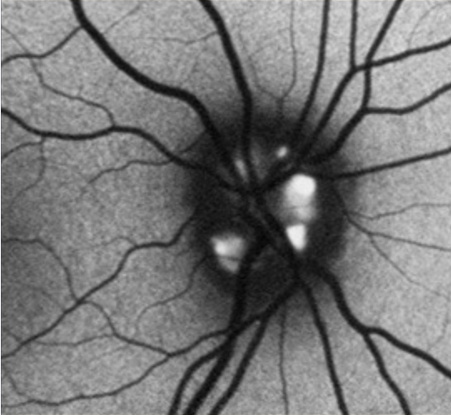
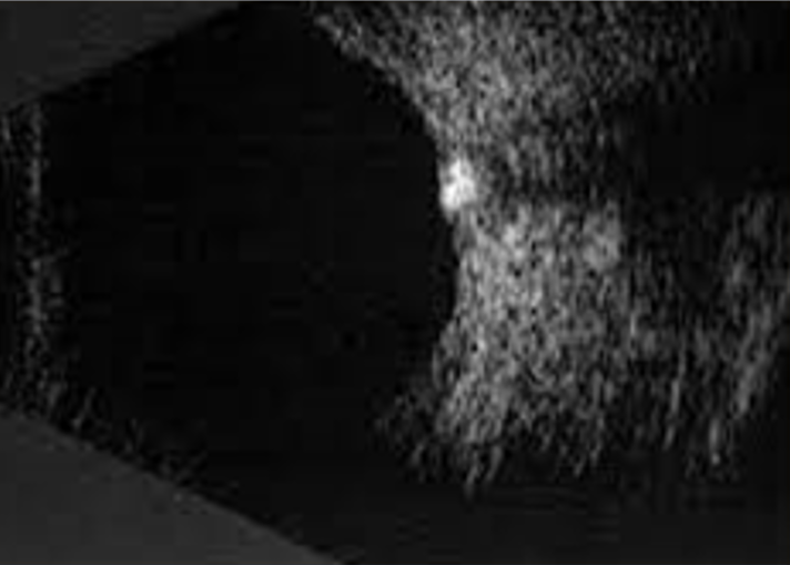
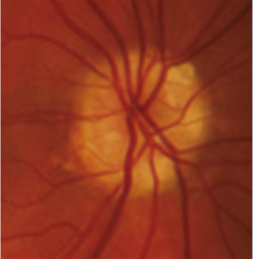
Optic disc drusen are accidental findings during an eye examination and represent acellular concretions predominantly made up of calcium, but can also contain amino and nucleic acids, mucopolysaccharides, and iron.(1)1 The primary pathogenesis of optic disc drusen remains unknown, with some hypothesising to be related to possibly inherited dysplasia of the optic disc with blood supply comprises, causing slowed axoplasmic flow and leading to the formation of calcific excrescences. (2,3) The optic disc drusen can be unilateral or bilateral, and according to literature, the vast majority are bilateral cases, and more often in females.(1) The reported prevalence of optic disc drusen is 1 to 500.(4)

Optic disc drusen can appear similar to papilloedema. This presentation is called pseudopapilloedema, and must be distinguished from actual optic disc oedema when examining these patients.

02

Symptoms & clinical signs

Optic disc drusen are primarily asymptomatic and a case of accidental finding during an eye examination. Approximately 9% of patients may present with visual obscurations.(5)

The optic disc drusen can be superficial, and in such cases, they give a “lumpy-bumpy” clinical appearance, or they can be located deeper within the optic nerve.

There are typical signs which should raise suspicion of optic disc drusen:

  • Elevated, often small, optic disc with indistinct and irregular disc margins.
  • Drusen are seen as round, white/yellow refractile bodies on the surface of the nerve or buried beneath it.
  • Anomalous vascular branching pattern (tortuosity, optociliary shunt vessels).
  • The nasal margin is the most common site of drusen.
  • Spontaneous venous pulsations.
  • The afferent pupillary defect if there is asymmetric nerve involvement.

The optic drusen can also cause visual-field defects. Those defects can be present in childhood and slowly progress over time.(6) There are two primary defects found in the optic disc drusen:

  • An arcuate defect in the inferonasal quadrant, secondary to direct compression or displacement of retinal nerve fibre layers by drusen.(7)
  • An enlarged blind spot due to pseudopapilloedema or leakage from the blood vessels.(8)

The visual defects are more often present in patients with superficial drusen.

Image 1. Obvious superficial optic disc drusen.
03

Diagnostic procedures

Slit lamp examination and colour fundus photography – superficial drusen are easily seen as lumps on the surface of the optic disc.

Fundus autofluorescence (FAF) – the optic disc drusen are highly auto-fluorescent, and FAF imaging is an excellent and helpful tool to recognise them. The imaging modality is used for the superficial optic disc drusen.

Image 2. Hyper auto-fluorescence of optic disc drusen.

Ultrasound is a great technique to recognise the buried (deeply located) optic disc drusen. It depends on the percentage of calcium within the drusen.

Image 3. Ultrasound of optic disc drusen. The ultrasound is a modality that can nicely present the optic disc drusen in case they contain calcium.

Optical coherence tomography provides the ability to detect optic disc drusen to the lamina cribrosa. It is an excellent tool to recognise and distinguish superficial from deep drusen.

Image 4. OCT image of the optic disc with superficial optic disc drusen. The arrow shows the hyperreflective lump within the optic disc representing a druse.
04

Associated retinal changes

Due to its location, the optic disc drusen may cause associated retinal changes, including retinal vasculature changes. Patients may present with tortuous retinal vasculature, vascular loops and/or optocilliary shunts. There are described case reports where patients developed central retinal artery occlusion (CRAO) and central retinal vein occlusion (CRVO) secondary to optic disc drusen. Uncommonly, patients with optic disc drusen can develop peripapillary choroidal neovascularisation (CNV), intraretinal flame-shaped haemorrhage, deep optic disc haemorrhage, or vitreous haemorrhage (rarely).(9,10)

05

How to distinguish papilloedema from pseudopapilloedema?

The appearance of optic disc drusen may lead to wrong suspicion of papilloedema, which is an urgent and potentially life-threatening condition. Therefore, it is essential to be familiar with specific clinical signs on distinguishing optic disc drusen-related pseudopapilloedema from the real swelling of the optic disc.


Congestion

Dilatation 

Retinal vessels 

True optic disc oedema 

Congestion of optic disc vasculature with a reddish hue of the disc

Dilatation of optic disc surface microvasculature/ flame-shaped haemorrhage 

Obscuration of the optic disc margin and retinal vessels at the edge of the disc

Pseudopapilloedema 

No congestion or reddish hue of the disc 

No dilatation of surface microvasculature of the optic disc

The retinal vessels are clearly visible

Table 1: Characteristics of papilloedema vs pseudopapilloedema

Image 5. The image represents the optic disc drusen. The nasal margins are obscured. However, there is the absence of a halo, and the retinal vessels are clearly visible.
Image 6. The image represents stage 3 papilloedema. Note the obscuration of leaving retinal vessels and a halo around the optic disc.
06

Management and treatment

There is no treatment warranted for optic disc drusen. Generally, the prognosis is good, and you should monitor your patient regularly. The potential retinal complications (CRAO, CRVO, peripapillary CNV) should be treated as required.

In the case of the progression of visual-field loss, there is no agreement on treatment. Some experts propose IOP-lowering eye drops.(11) Patients with optic disc drusen may have co-existing glaucoma, and monitoring glaucoma progression in such patients is challenging. You should refer those patients to an ophthalmologist so that they can be monitored.

07

References

1 Auw-Haedrich C, Staubach F, Witschel H. Optic disk drusen. Survey of ophthalmology. 2002 Nov 1;47(6):515-32.

2 Antcliff RJ, Spalton DJ. Are optic disc drusen inherited?. Ophthalmology. 1999 Jul 1;106(7):1278-81.

3 Sacks JG, O’Grady RB, Choromokos E, Leestma J. The pathogenesis of optic nerve drusen: A hypothesis. Archives of Ophthalmology. 1977 Mar 1;95(3):425-8.

4 Lorentzen SE. Drusen of the optic disc. A clinical and genetic study. Acta Ophthalmol (Suppl). 1966;90:1-81.

5 American Academy of Ophthalmology. Basic and Clinical Science Course. Neuro-Ophthalmology. The Patient With Decreased Vision: Classification and Management. Optic Disc Drusen. 2007-2008;129.

6 Sadun AA, Wang MY. Abnormalities of the optic disc. Handbook of clinical neurology. 2011 Jan 1;102:117-57.

7 Francois P. Hyaline verrucosites of the papilla. In Annals of Oculistics 1949 (Vol. 182, pp. 249-78).

8 Oliveira-Ferreira C, Leuzinger-Dias M, Tavares-Ferreira J, Faria O, Falcão-Reis F. The Relationship between Intraocular Pressure and Optic Nerve Structural and Functional Damage in Patients with Optic Nerve Head Drusen. Neuro-Ophthalmology. 2020 Sep 2;44(5):290-3.

9 Wessing A. Fluoreszenzangiographie der Retina: Lehrbuch und Atlas. Geleitwort von G. Meyer-Schwickerath.[Mit] 120 Abbildungen in 463 Einzeldarstellungen, 11 Tabellen. G. Thieme; 1968.

10 Auw-Haedrich C, Staubach F, Witschel H. Optic disk drusen. Survey of ophthalmology. 2002 Nov 1;47(6):515-32.

11 Pojda-Wilczek D, Wycisło-Gawron P. The effect of a decrease in intraocular pressure on optic nerve function in patients with optic nerve drusen. Ophthalmic Research. 2019;61(3):153-8.