Oct 4, 2021 • 30 minutes read

How to optimise glaucoma detection in everyday practice

Introduction

Written by
Topcon Healthcare

Glaucoma is an overarching term for different conditions that damage the optic nerve (known as glaucomatous optic neuropathy), cause visual field loss, and, if left untreated, result in blindness. Glaucoma is a dominant issue for optometrists. Not because we see lots of glaucoma patients, but because if we don’t detect suspected glaucoma and refer patients on, it is unlikely that anyone else will, due to the lack of symptoms in the vast majority of sufferers. Some cases are clear-cut, with obvious optic disc changes, arcuate visual field loss and raised intraocular pressure. However, many cases pose difficulties for ophthalmology colleagues as they are much more subtle. So, how are we supposed to refer correctly? On one hand, missing signs of glaucoma allow the glaucomatous optic neuropathy to progress along with the associated damage to vision. On the other hand, “false positive” referrals can cause a backlog in hospitals and unnecessary concerns for patients.

Due to the comprehensive research interest in glaucoma detection and treatment, new findings get published every month. These publications contain nuggets of information that optometrists should take on board and apply to their clinical practice to optimise the sensitivity and specificity of their portfolio of tests for early glaucoma detection. Admittedly, sifting through such a large amount of information is time-consuming for busy professionals. But help is at hand. This handbook highlights the most impactful research findings over the last few years, and they are directly applicable to your optometric practice. It provides five steps that are easy to implement and can make a big difference to you, your patients, and your ophthalmology colleagues.

This is a handbook by Catharine Chisholm and Chris Lee from Topcon Healthcare.


Chapters

Five steps to make your clinical practice reflect the latest best practice guidelines on detecting glaucoma

01
Know your glaucoma types

Open, closed, primary, secondary – and all the other terms you should have at the front of your mind.

Go to chapter
02
Be aware of the risks and act on it

A review of the factors that put an individual at greater risk of developing glaucoma

Go to chapter
03
Understand structural change in glaucoma

Tips for capturing quality OCT data and using it to support your clinical decision making

Go to chapter
04
Know how to optimise the assessment of visual function in glaucoma

Tips for efficient visual field testing while maintaining high sensitivity and specificity

Go to chapter
05
Consider the complete clinical picture

Piece together the jigsaw and be cautious about referring based on one abnormal finding alone.

Go to chapter
06
Want to learn more about glaucoma detection?

Go to chapter
01

Know your glaucoma types

Open, closed, primary, secondary – and all the other terms you should have at the front of your mind.

It is important to be aware of the different types of glaucoma and how they present themselves. Is the patient suffering from primary or secondary glaucoma? Open or closed-angle? And beyond the optic disc, what other ocular structures does glaucoma affect? Differentiation between the types of glaucoma supports your decisions about the appropriate referral speed and sometimes who to refer to. For example, is the patient having a closed-angle glaucoma attack and therefore needs an urgent referral to the emergency department?

Closed-angle glaucoma is more common than you think

The intraocular pressure of the eye depends on a balance between the production (ciliary processes) and drainage of aqueous humour. Drainage occurs primarily through the trabecular meshwork, located at the ‘corner’ of the anterior chamber angle, where the cornea meets the iris. While primary open-angle glaucoma shows the highest prevalence at around 74% of all glaucoma cases, around 20% are primary closed-angle glaucoma (PCAG). The angle is closed, because it is blocked by the peripheral iris. Closure or partial closure of the angle restricts the flow of the aqueous humour able to reach the trabecular meshwork and drain out of the eye

Closed-angle glaucoma

We are taught that primary closed-angle glaucoma is symptomatic, resulting in pain, photophobia, and blurred vision. Not to mention accompanied by signs of circumcorneal hyperaemia and a cloudy cornea. However, that is only true for the acute form. In contrast, chronic primary closed-angle glaucoma tends to have a similar clinical presentation to primary open-angle glaucoma – glaucomatous optic neuropathy, visual field loss and raised IOP, often limited to between 24-35mmHg. The patient tends to be asymptomatic or may report occasional episodes of discomfort and perhaps blurred vision associated with spikes in intraocular pressure. These episodes may only occur under specific environmental conditions that cause pupil dilation, such as driving at night. Growing evidence shows that chronic primary closed-angle glaucoma accounts for up to one in every five cases of closed-angle glaucoma. Patients can be misdiagnosed as having primary open-angle glaucoma because of the similarity in the clinical presentation.

You can differentiate between primary open-angle glaucoma and chronic primary closed-angle glaucoma by examining the anterior chamber angle and considering risk factors. In optometry practice, anterior segment OCT and van Herrick (slit lamp) methods are most commonly used to estimate the openness of the angle. Once a patient is referred to secondary care, ophthalmologists tend to use anterior segment OCT and/or gonioscopy, which is considered the gold standard. Risk factors for both acute and chronic forms of closed-angle glaucoma include Asian ethnicity, hyperopia and age. A closed or narrow angle is more likely to occur with increasing age due to the increased lens thickness pushing the peripheral iris forward. Treatment usually involves crystalline lens extraction.

Anterior segment OCT of an open anterior chamber angle

Primary or secondary?

While most glaucoma types classify as primary, a small proportion (around 6%) occur secondary to another condition. In most cases, the anterior chamber angle remains open, but aqueous humour drainage is impeded to a greater or lesser extent by different forms of debris such as inflammatory cells. As a result, the intraocular pressure rises. Here are some of the most common secondary glaucoma types:

  • Exfoliative glaucoma is a secondary condition related to Pseudoexfoliation syndrome. An age-related disease resulting in the deposition of white, flaky lens material onto the ciliary body and lens capsule. These deposits clog the trabecular meshwork and decrease aqueous outflow. The syndrome is more prevalent in Scandinavian populations and requires laser treatment or more invasive surgery to allow the aqueous humour to drain from the eye sufficiently.
  • Pigment dispersion syndrome can lead to pigmentary glaucoma. Iris pigment cells detach and float around in the aqueous humour. Over time, these pigment cells accumulate in the anterior chamber angle and clog the trabecular meshwork, causing the IOP to rise. Iris transillumination is a classic sign of pigment dispersion syndrome.
  • Steroids (whether taken systemically or applied to the eye) can cause intraocular pressure to spike in susceptible individuals. Steroid glaucoma is more common when the steroid is applied via an eye drop or injection into or around the eye and less common with steroid skin cream use. In most cases, the intraocular pressure returns to baseline levels when the steroid is withdrawn.
  • Neovascular glaucoma mainly occurs secondary to proliferative diabetic retinopathy or central vein retinal occlusion. Tiny new vessels develop over the iris and into the angle, which can obstruct aqueous humour outflow and lead to increased IOP.

You can often cure secondary glaucoma types by treating the underlying condition. If the treatment makes the intraocular pressure return to normal, further intervention may be unnecessary. Unfortunately, if axonal damage has already occurred, visual loss cannot be reversed. It should, however, stabilise once the pressure is normalised. To identify secondary causes, combine careful history taking with slit lamp and fundus/OCT examination.

Know the retinal layers affected by glaucoma

Optic neuropathy is the hallmark of glaucoma, and we are trained to look for an increase in the cup to disc ratio, notching, pallor, etc. However, glaucomatous damage is also visible elsewhere, and you can assess it using objective techniques.

The optic nerve is primarily composed of retinal ganglion cell (RGC) axons. Their cell bodies are located in the ganglion cell layer of the inner retina, and their axons form the retinal nerve fibre layer that traverses the retinal surface to the optic nerve head. Ageing naturally results in a loss of around 6000 retinal ganglion cells each year, which is seen on Optical Coherence Tomography (OCT) as a very gradual thinning of the retinal nerve fibre layer. Glaucoma causes an acceleration of this cell loss, and changes can be visible as a local thinning of the retinal nerve fibre and ganglion cell layers before noticeable changes occur at the optic nerve head. OCT scans the retina, applies a segmentation algorithm and provides highly repeatable thickness metrics for the retinal nerve fibre layer (RNFL) and ganglion cell layers (GCL). While the GCL and RNFL layers are composed of different parts of the same cells, evidence suggests that a separate evaluation can increase sensitivity and support earlier detection of glaucomatous damage.

Diagnostics in early detecting of glaucoma - Bharucha Kalyani et al.

Read more on the theory
Light passes through the inner retinal layers to reach the photoreceptors. The electrical signal is passed back up through a chain of retinal cells including the retinal ganglion cells and is carried by the ganglion cell axons to the brain, via the optic nerve.

Conclusion

We are all very familiar with the signs of primary open-angle glaucoma (POAG). But if you suspect glaucomatous optic neuropathy, don’t automatically assume that it is caused by the most common open-angle form. A quick check of the angle using either the AS-OCT or the van Herrick method can help identify patients who may be suffering from chronic closed-angle glaucoma. Generally, the condition can be successfully treated by removing the crystalline lens, but an important takeaway is that chronic closed-angle glaucoma is much more common than many optometrists realise.

Secondary glaucoma is rare, but you should nonetheless rule it out in patients with raised intraocular pressure. A careful history, combined with clinical tests including slit lamp, fundus exam and OCT) should be sufficient to rule out conditions that can cause a secondary rise in IOP, i.e., secondary glaucoma. Conversely, patients suffering from conditions such as uveitis, Pseudoexfoliation syndrome, pigment dispersion syndrome or retinal vein occlusion that can lead to secondary glaucoma should have their intraocular pressure checked, regardless of age.

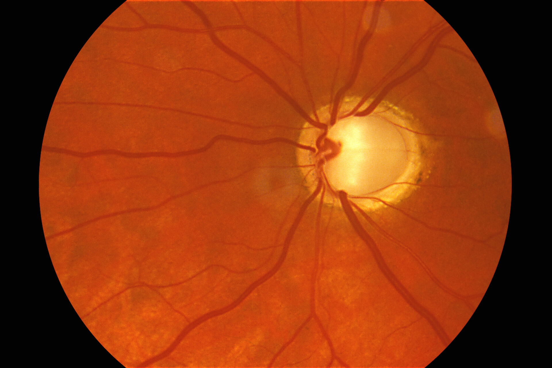
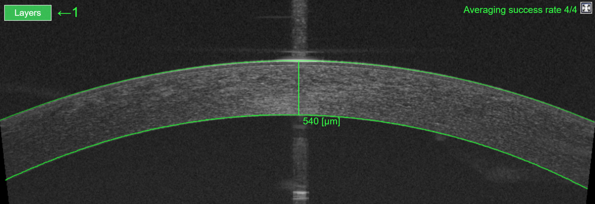
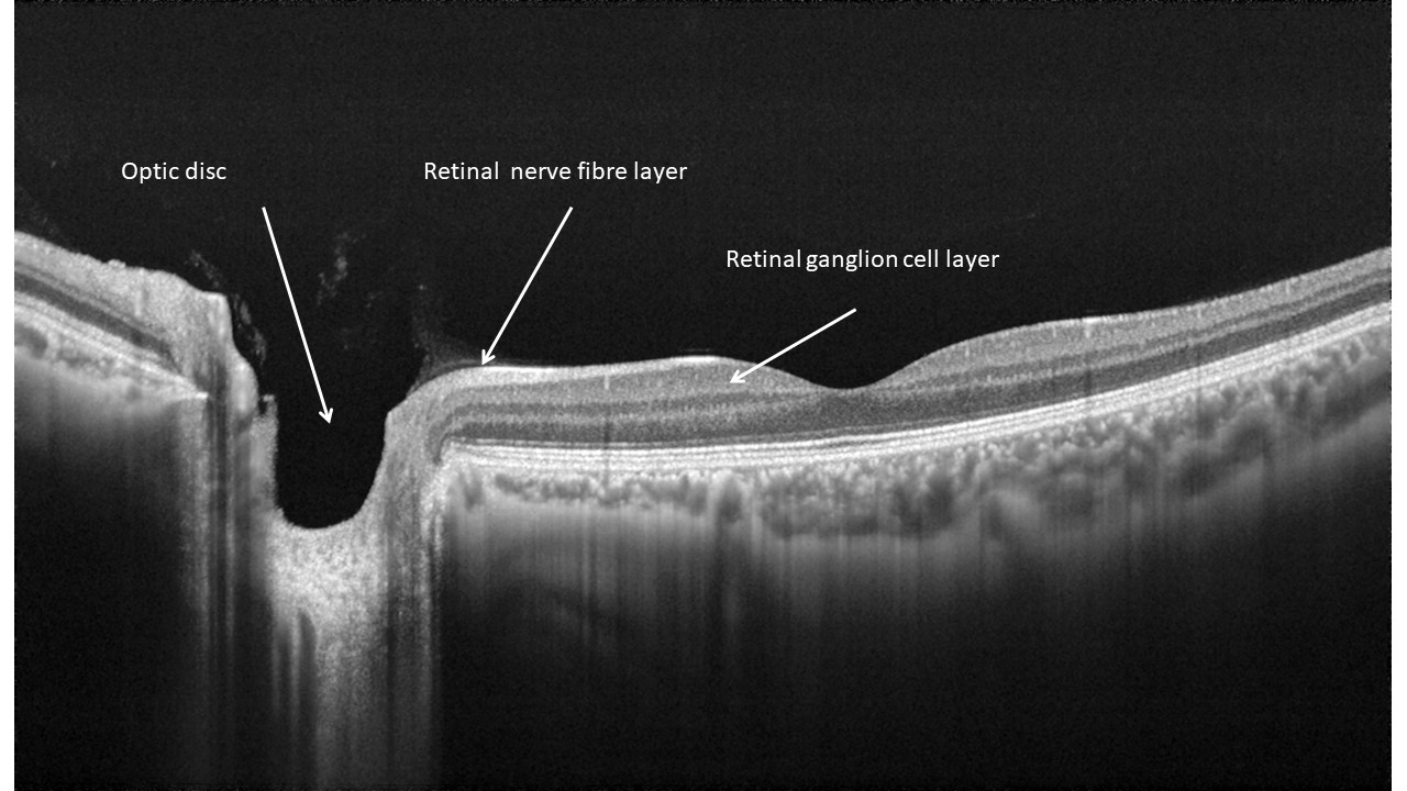
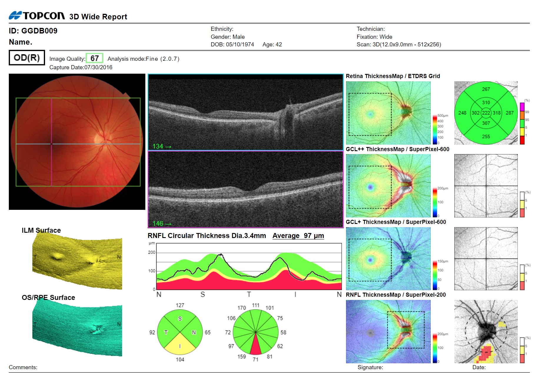
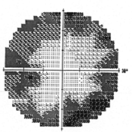
And finally, while many of us struggle to remember the numerous different layers of the retina, an OCT scan provides a rapid, automated analysis of the retinal layers affected by glaucoma (ganglion cell layer and retinal nerve fibre layer) without needing to get bogged down in the details of different layers. These measurements, which can be compared with reference data and/or monitored over time, provide additional information to complement optic nerve head findings that are infamously subjective. OCT metrics can lead to earlier detection of glaucomatous damage associated with asymptomatic glaucoma – i.e., primary open-angle glaucoma and chronic closed-angle glaucoma.

02

Be aware of the risks and act on it

A review of the factors that put an individual at greater risk of developing glaucoma

It is well-known that ethnicity, age and a family history of glaucoma increase the risk of a patient developing primary open-angle glaucoma. But population studies over the years have identified several other risk factors. The findings have resulted in a change in how professionals view raised intraocular pressure (IOP). Originally it was part of the definition of glaucoma, but that is no longer the case. Now it is considered a significant risk factor for developing primary open-angle glaucoma. Three theories of glaucomatous damage are relevant here because they help explain why glaucoma can occur without raised pressure and how some eyes can remain healthy despite increased intraocular pressure. A thorough clinical history and examination will reveal risk factors that should be considered as part of the whole clinical picture. This chapter explains some of the risk factors you may not be aware of.

Raised IOP increases the risk of glaucoma

Raised intraocular pressure is one of many risk factors for primary open-angle glaucoma. The higher the IOP, the greater the risk of glaucoma. An intraocular pressure greater than 22 mmHg without any glaucomatous damage classifies as ocular hypertension. Patients with this condition do not have glaucoma, but they need closer monitoring because of their increased risk of developing it. At the opposite end of the intraocular pressure scale, it is possible to have a pressure within the normal range (10-22 mmHg) associated with signs of glaucomatous damage. This classifies as normal-tension glaucoma.

Three theories of glaucomatous damage help explain why some patients have primary open-angle glaucoma despite having low intraocular pressure. And why others do not have glaucoma despite having high intraocular pressure.

  • Vascular theory: Retinal ganglion cells (RGC) are damaged by insufficient ocular blood flow due to either raised intraocular pressure or low perfusionof the capillaries that supply the optic nerve head. The resulting ischemia damages the retinal ganglion cell axons (the retinal nerve fibre layer – RNFL).  This theory helps explain the existence of normal tension glaucoma with low blood pressure and poor circulation associated with an increased risk of glaucoma.  But ischemic damage of the axons can also occur with high systemic blood pressure by causing haemorrhages – effectively creating a mini stroke within the optic nerve head, limiting blood supply to the axons. Read more here.
True colour fundus image of a patient with normal tension glaucoma, showing a disc haemorrhage. Courtesy of Professor Nicholas Rumney MSc Optom FCOptom DipTp(IP) Prof Cert Med RetMr
  • Mechanical theory: This theory proposes that the retinal ganglion cells are damaged by increased intraocular pressure, either directly or indirectly.
    Raised intraocular pressure exerts mechanical pressure and distorts the lamina cribrosa (the connective tissue structure within the optic nerve head). The retinal ganglion cell axons pass through the pores of this structure as they leave the globe. The axons may be damaged directly by the high pressure or indirectly as a result of lamina cribrosa distortion and compression. Read more here.
  • Biochemical theory: Various biochemical mechanisms may also play a role in glaucomatous neurodegeneration. These include damage by nitric oxide and oxygen free radicals (Ahmad 2016).

In reality, it is likely that the pathogenesis of glaucoma is multifactorial and varies between patients. For any glaucoma patient, one, two or all three of the above theories can help explain their glaucoma pathogenesis. Understanding this better may lead to improvements in glaucoma treatment in the future.  For now, however, the only real treatment we have for primary open-angle glaucoma is eye drops or procedures (lasers/surgery) to lower the intraocular pressure.

Corneal consideration

Do you remember when you were learning about the measurement of intraocular pressure? The applanation tonometry formula is based on the assumption that the eye is a dry, thin-walled sphere. Well, you will probably have gathered by now that it isn’t dry and it isn’t a thin-walled sphere. Some corneas are easier to deform than others, potentially leading to an underestimation of intraocular pressure. This ocular hypertensive study shows that the risk of developing primary open-angle glaucoma is inversely correlated with central corneal thickness. In addition to the measurement artefact associated with the fact that the eye is not a dry, thin-walled sphere, the researchers could not rule out additional biomechanical reasons why subjects with a thinner cornea are at greater risk of glaucoma. They discovered that participants with a corneal thickness of 555 µm or less have a three times greater risk of developing primary open-angle glaucoma than participants, who have a corneal thickness of more than 588µm.

Measurement of corneal thickness based on an anterior segment OCT scan

Also, you must remember that some patients will have a thinner and more easily deformable cornea due to pathologies such as keratoconus (assuming no cross-linking treatment) or corneal refractive surgery such as LASIK. Your threshold for concern about measured IOP should be adjusted accordingly. For example, a reading of 21mmHg may indicate an IOP of 22.5 or even 24mmHg, depending on how thin the cornea is. This article helps describe how corneal thickness can affect the reliability of IOP measurement.

Keeping this in mind, make sure you ask about refractive surgery during history taking and consider measuring corneal thickness. It will help to identify patients who you should monitor more closely. Anterior segment OCT is a fast way to measure the central corneal thickness (CCT) but air-puff tonometers – such as the TRK-2P – also provide a quick measurement of CCT and automatically adjust intraocular pressure values for corneal thickness.

Some air puff tonometers measure central corneal thickness alongside intraocular pressure

Myope or hyperope?

According to a study by Michael W. Marcus and colleagues, medium and high myopes are up to 60% more likely to develop primary open-angle glaucoma than an emmetrope. That is one of the many reasons why clinicians should offer myopia management to slow the development of myopia in children. The greater risk of glaucoma is probably multifactorial; axial elongation of the globe due to myopia certainly plays a role. The retina and optic nerve are under more biomechanical stress. And the characteristics of the myopic eye – a large optic disc, peripapillary atrophy and a thin retina – make it harder to differentiate early glaucoma from a healthy eye. The retinal nerve fibre layer thickness of a high myope tends to lie at the thin end of the reference population. Therefore, myopes are more likely to show ‘red disease’ on OCT (thickness measurements that dip into the concerning red area of the reference database but are nevertheless normal for that individual).

So, is OCT still useful in myopes? Absolutely! But progression data may be better than OCT data from a single visit. Therefore, you may want to perform a ‘wellness’ scan on your myopic patients under 40 years, as it is useful to have previous scans to compare with. Add ocular parameters into the patient profile in your OCT software, such as axial length, or, at least, add the refractive error details so that thickness measurements are corrected for the effect of ocular magnification.

At the other end of the scale, don’t forget that hyperopes tend to have a shorter than average axial length and a shallow anterior chamber. That puts them at a greater risk of suffering from primary closed-angle glaucoma. A quick anterior segment OCT scan or van Herrick will provide information about whether the anterior chamber angle is open/closed or narrow and therefore at risk of closure.

Conclusion

Be aware of the risk factors for primary open-angle glaucoma. A careful history can identify low or high systemic blood pressure – both of which increase the risk of primary open-angle glaucoma based on the vascular theory of glaucomatous damage. It is worth remembering that there is a balance between the measured intraocular pressure and the pressure that the ocular structures can withstand. One person’s eye may be able to withstand quite a high pressure without any damage occurring, whereas another person may suffer from glaucomatous damage at a pressure well within the ‘normal’ range. Make sure you are familiar with the definitions of normal-tension and high-tension glaucoma, along with ocular hypertension.

A measurement of intraocular pressure enables you to identify patients with raised values, who are at greater risk of developing glaucoma. Additional measurements such as refractive error and corneal thickness can help you further refine the level of glaucoma risk for an individual. The higher the refractive error, the greater the risk, with myopes at greater risk of developing primary open-angle glaucoma and hyperopes at greater risk of developing primary closed-angle glaucoma.

Use an air puff tonometer that incorporates corneal thickness measurement such as the TRK-2P or capture an anterior segment scan on your OCT to assess the corneal thickness and identify patients with thinner corneas who are at greater risk of primary closed-angle glaucoma. Since primary open-angle glaucoma is generally asymptomatic, early diagnosis of glaucoma is critical for long term visual prognosis. Anything we can do to detect glaucoma earlier should be done, including performing a more detailed and frequent examination of those with a higher risk of glaucoma.

03

Understand structural change in glaucoma

Tips for capturing quality OCT data and using it to support your clinical decision making

Optical Coherence Tomography (OCT) is widely used in ophthalmology across the majority of specialities, including glaucoma. Over the last 5-10 years, it has become much more common in primary eye care settings because it provides good support for referral decisions. The automated layer segmentation provides thickness measurements, which are invaluable in the detection and management of glaucoma (in addition to generating a cross-sectional view of the posterior segment and other parts of the eye). OCT allows the clinician to examine the eye for the structural effects of glaucoma, which often arise before any loss of the visual field.

The key structures to image in glaucoma are the optic disc, retinal nerve fibre layer and the ganglion cell layer

The key retinal layers of interest concerning glaucoma are the retinal nerve fibre layer (RNFL) and the ganglion cell layer (GCL). The OCT analysis uses the following definitions for each layer:

  • Retinal nerve fibre layer: Tissue thickness between the inner limiting membrane and the boundary between the RNFL and the GCL.
  • GCL+ is between the RNFL/GCL boundary and the inner plexiform layer/inner nuclear layer boundary.
  • GCL++ is effectively the RNFL and GCL+ layers added together (inner limiting membrane to inner plexiform layer/inner nuclear layer boundary).

Go wide (and dense)

The scan speed of modern OCT devices is around 50,000 A-scans per second, with a reachable 100,000 A-scans per second for swept-source OCT devices. These speeds allow dense data sets to be collected and greatly reduce the chance of missing information that falls between scan lines. The Topcon 3D OCT-1 Maestro2 offers a 12x9mm scan pattern covering the disc and macular regions in a single scan. This consists of 128 B scans, each containing 512 A-scans. That’s a very dense dataset. While it is possible to scan the macula and the disc separately, a wide field scan has significant benefits, both in terms of capture time and when it comes to reviewing the data. Not only is it quicker to review a single report but it makes more sense clinically since both macular and disc areas are affected by glaucoma and damage tends to be continuous between the two regions.

Perform quality control

OCT thickness measurements are very repeatable but always dependent on the quality of the scan. Before you review the OCT results, check the quality score. There are various reasons for a low scan quality: Cataract reducing the amount of light reaching the retina. Dry eye distorting the light entering the eye. Poor patient instruction. It is important to ask all patients to take a few good blinks immediately prior to the OCT capture. Clear instruction is even more important with patients with a poor-quality tear film, and those with very dry eyes may benefit from the instillation of a drop of artificial tears immediately prior to capture to create a smooth refracting surface for the OCT beam entering and leaving the eye.

In addition to scan quality, you should also check for artefacts associated with eye movements or blinks. These artefacts show up in the scan preview as black lines or jagged blood vessels. When they are present, it is worth repeating the scan to see if the quality can be improved by asking the patient to keep their eyes wide open during capture and reminding them throughout capture to keep looking at the green target.

Click here for a capture-optimisation training or visit Topcon Healthcare University online for a range of OCT-related courses.

Example of a poor-quality OCT scan due to eye movement during capture, indicated by the jagged blood vessels and horizontal band.

How does the patient compare in the reference database?

All commercially available OCTs include a database based on a large reference population. The option allows RNFL, GCL+ and GCL++ thickness measurements for an individual patient, which you can compare with the measurements expected for the patient´s age group, defined by the reference database population. For any layer and retinal location, the reference population will demonstrate a range of thickness measurements. They form a bell-shaped normal distribution. The majority of patients have thickness measurements that correspond to the central body of the bell curve (middle and either side), while some have thicker or thinner values that fall into the curve´s two tails. Thickness values falling within the main central body of the normal distribution curve are colour-coded as green on OCT reports. Around 4% of the reference population will show thinner measurements in the tail of the distribution and well outside the main central body of the distribution. Thickness values in this region are colour-coded yellow or orange. A further 1% of the population will show very thin values at the tail end that are colour-coded red.

OCT reports present the measured retinal nerve fibre layer thickness around the optic disc, colour-coded to compare with the reference data. The thickness data is averaged and displayed over four quadrants and also 12 clock segments. If one or more segments have yellow or red thickness measurements, it indicates that the retinal nerve fibre layer thickness corresponds to the thin tail of the reference database distribution. Consequently, there is a higher suspicion of glaucoma. Since glaucoma rarely develops symmetrically, yellow and red segments for one eye but not the other eye increase the level of suspicion further. However, interpret the colour-coded comparison with the reference data with caution. The presence of yellow or red segments does not definitively indicate glaucoma. They merely increase the chance of glaucoma being present. Some “normal” patients without glaucoma have a naturally thin retinal nerve fibre layer all their lives, corresponding to the yellow or red tails. It may be because they have a longer eye and hence a thinner retina.

There are also variations on normal anatomy, including tilted discs, large optic discs, eyes with a large angle between the major retinal arcades. All these normal variations can lead to yellow or red segments on the OCT report, without glaucoma being present. Conversely, some patients with glaucoma will have retinal nerve fibre layer thickness measurements well within the green range of the reference database and yet be diagnosed with glaucoma. That can occur if the patient started life with a particularly thick retinal nerve fibre layer. The 3D OCT-1 Maestro2 has the advantage of simultaneously capturing a colour fundus image, which is pinpoint registered to the OCT scan. The fundus image provides information about wedge defects or confounding retinal disease along with the disc appearance, including variations on normal anatomy.

This patient’s 3D wide scan report shows a yellow, inferior quadrant and a single red inferior clock segment, indicating a thin retinal nerve fibre layer at this location. The report for the left eye and other clinical findings and risk factors should be examined before assuming this is glaucoma.

Conclusion

OCT is already providing valuable insight into the detection and management of glaucoma and retinal disease. Capturing a wide field OCT scan covering a 12x9mm area provides an analysis of the retinal layers affected by glaucoma over both the disc and macular regions. The layers of interest are the retinal nerve fibre layer (around the optic disc and generally across the posterior pole) and the ganglion cell layer (around the macula). You can compare thickness data for both layers with a reference population but be careful to ensure the quality of scans captured since it impacts the accuracy of the thickness measurements.

Suspicious OCT findings indicated by yellow or red colour-coding of thickness measurements – compared to the reference database – do not necessarily indicate glaucoma. It is important to rule out other possible causes, such as tilted discs or high myopia. You should always interpret OCT findings in the context of the complete clinical picture, including the disc and fundus appearance, history, risk factors, and other clinical findings. It is rare to find a suspicious OCT report without there also being risk factors for glaucoma or some other clinical findings that raise suspicion of glaucoma.

With the rapid spread of OCT use in primary eye care, it won’t be long before we can reap additional benefits based on repeat scans on the same patient. Historical data allows for progression analysis, providing an extra tool for the detection of glaucoma but also other neurodegenerative conditions that can cause RNFL loss such as Alzheimer’s disease and multiple sclerosis. In the future, OCT Angiography (OCTA) is likely to become more widely used in glaucoma detection and management. It can support clinical decision making in tricky cases by imaging the Radial Peripapillary Capillaries (RPC) to look for vascular defects that may pre-empt visible retinal nerve fibre layer damage. Read more.

Vascular density map based on OCT Angiography, showing arcuate loss of radial peripapillary capillaries in glaucoma
04

Know how to optimise the assessment of visual function in glaucoma

Tips for efficient visual field testing while maintaining high sensitivity and specificity

Visual field loss is not something that patients report. The reason is that primary open-angle glaucoma develops slowly, impacts one eye before the other, and does not tend to affect central vision until the late stages of the disease. Although glaucoma can affect many aspects of vision (including paracentral contrast vision, colour vision, and motion perception), perimetry is the easiest and most commonly used method to examine visual function.

The Goldilocks’ test: Not too long, not too short, but just right

When screening visual fields for signs of glaucoma, you must choose the right test. As optometrists, we are interested in monitoring the progression of visual field loss, and therefore complete threshold testing is valuable. The metrics allow progression analysis to support treatment decisions. But complete threshold tests are time-consuming and stressful for patients, and therefore not practical for glaucoma screening. When it comes to visual field testing, the reliability of the data is dependent on a fine balance between the thoroughness of the test and the duration of the test. Testing more points will increase sensitivity to early glaucomatous field loss, but longer-lasting tests result in fatigue, variations in fixation and less reliable patient responses (ultimately increasing false positives).

Grey scale plot of a visual field test showing the characteristic ‘clover-leaf’ pattern seen in patients who are getting tired during the long test. This is not a visual field defect.

An efficient visual field screening test is required to avoid a significant impact on clinical workflow. Conventional screening tests present a suprathreshold target at each particular visual field location. This stimulus is slightly brighter than the one a patient of that age would expect to see based on reference data.

Modern screening tests such as the Henson Smart Supra take screening to a new level. Test points are located in the visual field areas most susceptible to early glaucomatous damage rather than a regular grid pattern. Also, the test offers the flexibility to retest points, investigate additional ones in the same area or extend with an additional test pattern. You can thus avoid performing a whole additional test of, for example, the central 10 degrees. It takes less than 1 minute to test one eye of a healthy patient while offering high sensitivity and specificity – the Goldilocks test.

Correct the refractive error

Visual field tests generally have a working distance of 25-30cm. Visual field thresholds are affected by the presence of an uncorrected refractive error, so it is important to correct it. Uncorrected refractive error causes generalised depression of the visual field, which shows up on the mean deviation map and can mask early visual field loss.

The refractive correction worn for near vision is the most suitable, but patients who wear bifocals or multifocals will need to view the test through an auxiliary lens to avoid suppressing the upper portion of the visual field. Make sure the auxiliary lens is well-positioned to avoid generating rim artefacts. If delegating visual field testing to your support staff, choose a visual field screener that auto-calculates the correction needed based on the patient’s spectacle prescription. That will help minimise measurement errors due to refractive blur.

When to perform visual fields

Although test algorithms like Smart Supra make wide-spread visual field testing a reality, visual fields are usually performed when there is a reason to be suspicious. For example, in case of a suspicious optic disc appearance, thinning of the RNFL or GCL on the OCT report, or the presence of risk factors such as raised IOP, thin cornea, family history of glaucoma etc.

Some OCTs can generate a Hood report from a 3D wide scan. The Hood report is designed by U.S. professor Don Hood from Columbia University to support early glaucoma detection. It incorporates OCT probability maps and highlights areas of the retinal nerve fibre layer or ganglion cell layer with a high probability of being abnormal. Helpfully, the 24° and 10° visual field test locations are superimposed over the probability maps. The location of suspicious areas on the probability maps can help you decide which visual field test to perform. If the suspicious areas coincide with some of the 24-2 test points, then a visual field test over 24° is a natural choice. If, however, the suspicious areas on the OCT probability map are limited to the central area, a 10° visual field test is recommended, because a test over 24° is not sensitive to early central visual field loss.

Remember that structural (OCT) and functional (visual field) losses do not necessarily occur in parallel. OCT is very sensitive, picking up thinning of retinal layers of just a few microns. Perimetry, in comparison, is a very subjective and crude measure of target detection over a grid pattern that may or may not coincide with the area of damage. In addition, an estimation is that between 40-50% of retinal nerve fibres need to be lost before a visual field defect becomes apparent. Hence, you would expect early structural glaucomatous damage to show on an OCT before a functional loss can be detected using visual fields.

Perimetry looks for functional loss related to glaucoma

Conclusion

If you are going to assess visual fields, do it right!  Choose a test optimised for glaucoma screening that will not significantly extend your examination time. Make sure that the patient is fresh and not tired when starting a test, and bring them back another day if necessary. Keep them engaged throughout the test. Choose a smart screening test that keeps the test time short but has good sensitivity and specificity, such as the extendable Smart Supra test. Implementing short test times makes it more realistic to offer visual field testing to all patients with a higher risk of glaucoma.  Remember that functional damage detectable by subjective visual field tests tends to occur after the structural glaucoma damage that is revealed through an OCT scanning. The Hood report provides a method to link structural and functional damage, and it supports the choice of the visual field test area based on the location of areas of suspicion on OCT.

05

Consider the complete clinical picture

Piece together the jigsaw and be cautious about referring based on one abnormal finding alone.

An integral part of optimising glaucoma detection is to consider the complete clinical picture. Look at risk factors based on the history, symptoms, and additional clinical measurements such as corneal thickness. Alongside, you should consider the colour fundus image, OCT analysis and perimetry results.

Make use of hanging protocols

Viewing clinical data on one screen enhances clinical decision making and improves efficiency. Software such as Harmony RS offers a hanging protocol function, allowing you to decide how you want clinical information presented to you. Alternatively, you can select one of the default hanging protocols, such as colour fundus image/OCT/visual field report, which will be presented side-by-side on the screen for any visit you select from the timeline. The platform is vendor-neutral, so you can freely choose devices to connect. The morph function provides an easy way to compare visual field or OCT reports between visits.

Hanging protocols allow you to view and interrogate the key clinical findings from a particular patient visit, on one screen. This example shows the colour fundus alongside the associated OCT B-scans and the visual field plot from the same clinical visit.

Do not refer based on one abnormal finding

Do not be tempted to refer based on one apparent abnormal finding such as raised pressure or an OCT scan with circumpapillary RNFL sectors that are red (thin) compared to the reference database. In such cases, additional tests or a second opinion from a colleague or an ophthalmologist can help you decide whether to refer or not. Look out for normal anatomical variations, particularly in higher myopes. Be suspicious if similar disc or OCT findings occur for both eyes – glaucoma is rarely – if ever – symmetrical in its development. Large optic discs, tilted disc, a large angle between the disc and fovea, or a large angle between the retinal arcades are all anatomical reasons why the OCT or disc metrics for a patient may not be reflected within the reference population, resulting in a false positive.

Use local referral criteria

Familiarise yourself with your national or local referral criteria for suspected glaucoma. Which patients do the ophthalmologists want to see, and what information do they want to receive from you? OCT and visual field reports along with pressure measurement can be shared through the referral mechanism in software such as Harmony RS. You may have the option of seeking a second opinion through Harmony RS, before deciding whether or not to refer. Sharing information to avoid the patient having to repeat tests is generally appreciated by all involved.

Conclusion

Going forward, communication between primary and secondary eye care needs to increase, if we want to provide an efficient system for managing the growing and ageing population. We already see technologies designed to support shared care and delegation to primary care, the prime example being Artificial Intelligence (AI) technology. For example, you can plug Thirona AI technology into Harmony RS to provide automated analysis of the fundus image for age-related macular degeneration, diabetic retinopathy or glaucoma. We are likely to see more AI technology in the future.

06

Want to learn more about glaucoma detection?

Register for Topcon Healthcare University online and access free e-learning courses that you can work through to grow your knowledge and confidence with glaucoma detection and OCT. Topcon has a wide range of courses available – including OCT interpretation, case presentations from doctors, OCT in glaucoma, understanding the Hood report, and OCT capture.

If you want to become a real expert in OCT and are willing to invest more time, consider taking one of the training modules from GREG (Gloucester Retinal Education Group), a world-renowned centre for retinal imaging education. See more here.