Sep 22, 2021

Chapter 1: Uvea

Introduction

Written by
Viewpoint

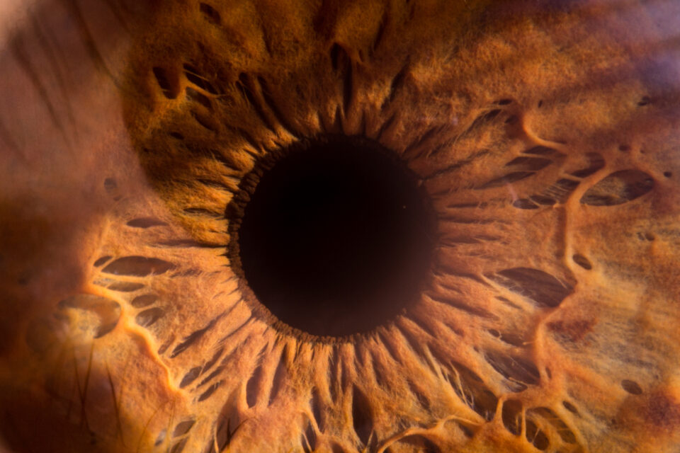
The uvea is the layer of the eye that’s between the retina and sclera and is the main blood supply for many ocular structures. It is composed of the iris, ciliary body, and choroid. This chapter covers the following diseases affecting the uvea:

  • Posterior uveitis
  • Toxoplasmosis
  • Ocular sarcoidosis
  • Choroidal nevus
  • Choroidal melanoma

Click on one of the cards below to read more about the specific eye condition.


Chapters
01
Posterior uveitis

Referral: Urgent*

Go to chapter
02
Toxoplasmosis

Referral: Urgent if active*

Go to chapter
03
Ocular sarcoidosis

Referral: Urgent if active*

Go to chapter
04
Choroidal nevus

Referral: Depending on risk factors*

Go to chapter
05
Choroidal melanoma

Referral: Urgent to normal depending on characteristics*

Go to chapter
01

Posterior uveitis

Referral: Urgent*

Uveitis is a general term describing a group of inflammatory diseases that produce swelling and destroy eye tissues.

In many cases, the cause of uveitis is unknown, but it may be caused by:

  • an attack from the body’s own immune system
  • infections or tumours occurring within the eye or in other parts of the body
  • bruises to the eye
  • toxins that may penetrate the eyeball

The disease will cause the following symptoms:

  • decreased vision
  • pain
  • light sensitivity
  • increased floaters
Posterior Uveitis
Image 1: Choroiditis
These images were originally published in the Retina Image Bank. Ivan R. Batlle, MD. Retina Image Bank. Year 2012; Image Numbers 761 and 762. © the American Society of Retina Specialists.
Image 2: Posterior uveitis with vitreous inflammation
This image was originally published in the Retina Image Bank. Dr Purva Patwari, Patwari Retina Center. Retina Image Bank. Year 2018; Image Number 28275. © the American Society of Retina Specialists.
02

Toxoplasmosis

Referral: Urgent if active*

Toxoplasmosis is a disease that results from infection with the toxoplasma gondii parasite – one of the world's most common parasites.

  • Infection usually occurs by eating undercooked contaminated meat, exposure from infected cat faeces, or mother-to-child transmission during pregnancy.
  • Once the parasite reaches the retina, it proliferates within host cells followed by rupture of the host cells, and invasion into neighbouring cells to make primary lesions.
  • Sometimes the restricted parasite by the host immunity in the first scar is activated to infect another lesion nearby the scar.
  • Blurred vision is the main complaint of ocular toxoplasmic patients and can be diagnosed by the detection of antibodies or parasite DNA.
  • Most cases of toxoplasmosis will be inactive and don’t require follow-up or referral.
Image 3: Congenital toxoplasmic scar
Image 4: Reactivation of the lesion 30 years after birth
03

Ocular sarcoidosis

Referral: Urgent if active*

Sarcoidosis is a chronic granulomatous disease of unknown aetiology characterised by the presence of non-necrotizing granulomata.

Image 5: Ocular sarcoidosis causes typical “candle wax” lesions in the central and midperipheral parts of the choroid
  • The reported incidence of sarcoidosis varies from 6 – 70 per 100,000 people per year.
  • Sarcoidosis typically affects the lungs and hilar lymph nodes, although any organ system may be affected, which may pose a diagnostic challenge.
  • Ocular involvement is a well-recognised extra thoracic complication of systemic sarcoidosis. The reported prevalence of ocular disease among patients with systemic sarcoidosis varies from 12 to 50%.
  • Uveitis is the most common ocular disease, although any eye structure may be affected. It’s important to recognise uveitis timely as it, depending on the pathology, will affect some or all parts of the eye and cause irreversible damage.
Image 6: Peripheral anterior synechiae in sarcoidosis – gonioscopic view
04

Choroidal nevus

Referral: Depending on risk factors*

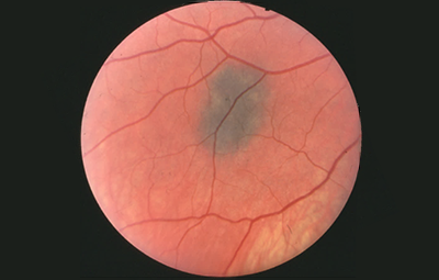
Choroidal nevus is the most common intraocular tumor, occurring in about 7% of adults. It is a benign melanocytic tumor.

  • Although it may be congenital, choroidal nevus is not typically observed in small children. Usually, it is first detected in adulthood on a routine eye examination.
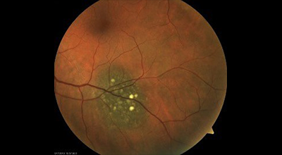
  • Choroidal nevi are slate-grey and relatively flat (< 2 mm thickness) and may show overlying drusen or lipofuscin. (See image 10)
  • Nevus almost never causes any symptoms, unless it is under the macula.
  • Very rarely, a choroidal nevus may leak fluid or be linked to the growth of abnormal blood vessels. These events can lead to retinal detachment, flashing lights, and vision loss.
  • Choroidal nevi have a risk, like with skin moles, to evolve into melanoma – therefore, nevi must always be monitored. Any change should raise the suspicion of malignant transformation. (See image 11)
Image 7: A choroidal nevus
Image 8: A choroidal nevus with overlying lipofuscin
Image 9: Tumor touching the disc contains a greater risk to evolve into melanoma
05

Choroidal melanoma

Referral: Urgent to normal depending on characteristics*

The annual malignant transformation rate of choroidal nevi in Caucasians is estimated to be one in 8845, this drops to one in 3664 for ages 80-84 years (Chien et al., 2017).

  • There are a number of risk factors for such melanoma transformation, the most important of which appears to be an initial thickness of more than 2 mm.
  • Carol Shields at Wills Eye Hospital (USA) has done a lot of work in this area. She identifies five factors associated with the risk of growth of small choroidal lesions:
    1. Tumour thickness greater than 2.0 mm
    2. Subretinal fluid
    3. Visual symptoms
    4. Orange pigment
    5. Posterior tumour margin touching the disc.
  • Nevi with three or more risk factors demonstrate a malignant transformation in 50% of cases over 5 years (Chien et al., 2017).

You can easily remember these five factors with the following mnemonic:

TFind Small Ocular Melanoma

translates into

Thickness, Fluid, Symptoms, Orange pigment, Optic Disc Margin
Image 10: Choroidal melanoma
This image was originally published in the Retina Image Bank. Jason S. Calhoun, Department of Ophthalmology, Mayo Clinic Jacksonville, Florida. Year 2013; Image Number 10386. © the American Society of Retina Specialists.
Image 11: Choroidal melanoma with pigment
This image was originally published in the Retina Image Bank. Jason S. Calhoun, Department of Ophthalmology, Mayo Clinic Jacksonville, Florida. Year 2013; Image Number 7799. © the American Society of Retina Specialists.

*Referral advice

The referral advice in the atlas should be used as a general guideline. The Optometry Atlas does not establish a standard of optometric care and specific outcomes are not guaranteed. Please read the full details here.