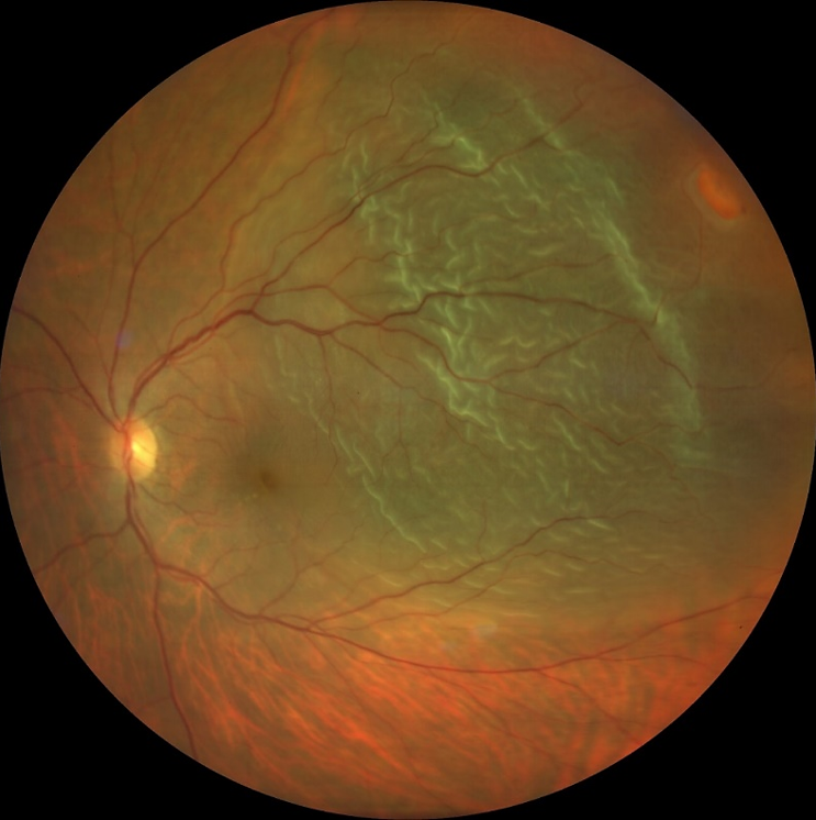
- Lattice degeneration is considered the most important peripheral retinal degeneration process that predisposes to a rhegmatogenous retinal detachment. (See image 1)
- Normally, the retinal pigment epithelium is able to maintain adhesion with the overlying neurosensory retina through a variety of mechanisms: active transport of subretinal fluid and inter-digitation of outer segments and the retinal pigment epithelium microvilli.
- Retinal detachment occurs when subretinal fluid accumulates between the neurosensory retina and the retinal pigment epithelium. This process can occur in three ways:
- a break in the retina allowing vitreous to directly enter the subretinal space. This is called a rhegmatogenous retinal detachment (often due to retinal tears associated with posterior vitreous detachment or trauma). (See image 2)
- proliferative membranes on the surface of the retina or vitreous -membranes can pull on the neurosensory retina causing physical separation between the neurosensory retina and retinal pigment epithelium. This is called a traction retinal detachment.
- accumulation of subretinal fluid due to inflammatory mediators or exudation of fluid from a mass lesion. This is known as a serous or exudative retinal detachment.

Sep 22, 2021
Chapter 3: Vitreoretinal interface abnormalities
Introduction
The vitreoretinal interface is a term for the connection between the vitreous and the retina (inner limiting membrane). This chapter covers retinal detachment.
Chapters
01
Retinal detachment
Referral: Urgent*
Retainal detachment is a sight-threatening condition with an incidence of 1 in 10,000 people.

Image 1: Retinal tear with Lattice degeneration
Courtesy of Zeiss
Image 2: A retinal detachment caused by a superotemporal horseshoe tear
Courtesy of Zeiss: Jean François Korobelnik, M.D. Universite de Bordeaux*Referral advice
The referral advice in the atlas should be used as a general guideline. The Optometry Atlas does not establish a standard of optometric care and specific outcomes are not guaranteed. Please read the full details here.


