Search results
You searched for “Imaging/OCT“
-
![]() Guide
GuideHow to optimise glaucoma detection in everyday practice
Five steps to make your clinical practice reflect the latest best practice guidelines on detecting glaucoma -
![]() CET module
CET moduleEssentials of OCT
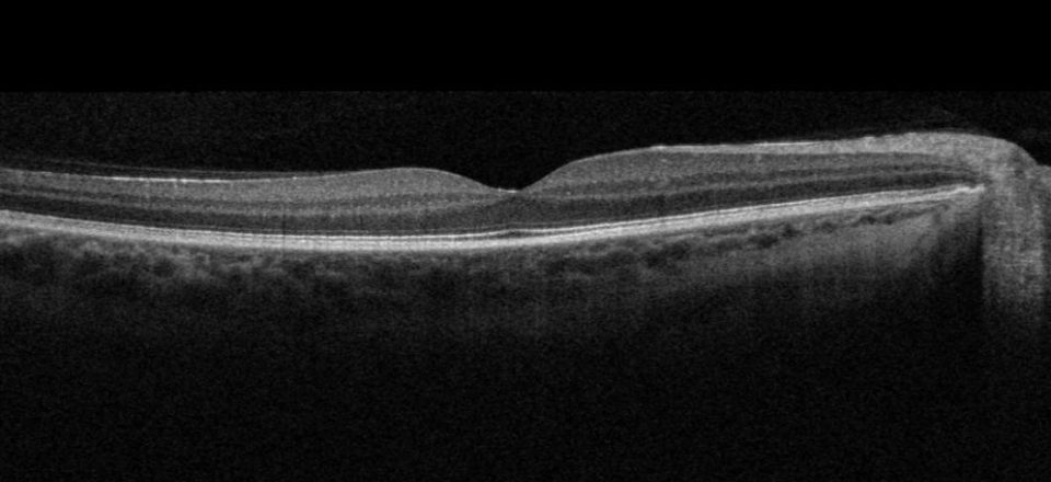
OCT is a medical imaging technique that is now comprehensively used by ophthalmologists and optometrists to acquire high resolution images of the anterior segment and retina. -
![]() CET module
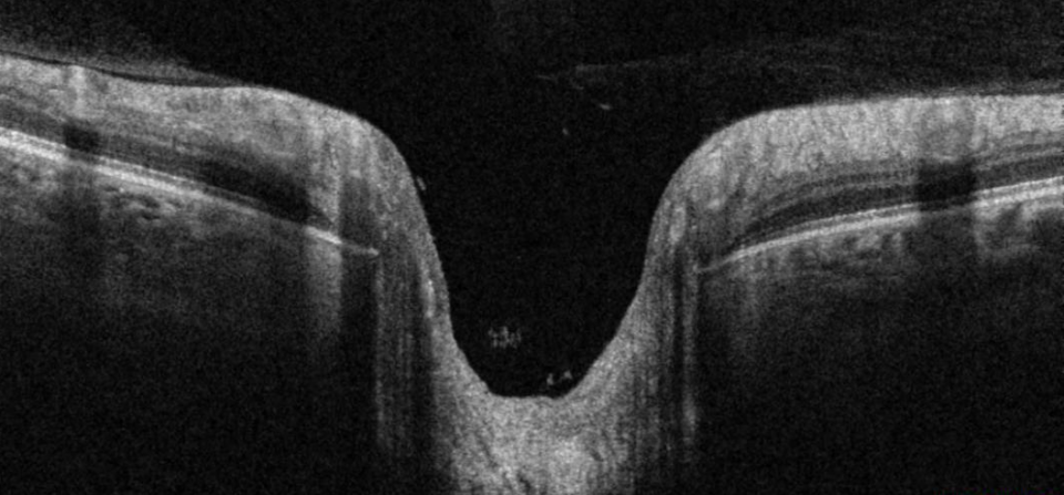
CET moduleOCT in glaucoma
This article will examine what OCT can tell us about the structural changes associated with glaucoma and consider how OCT can help in the diagnosis and management of this condition. -
![]() CET module
CET moduleOCT and medical Retina
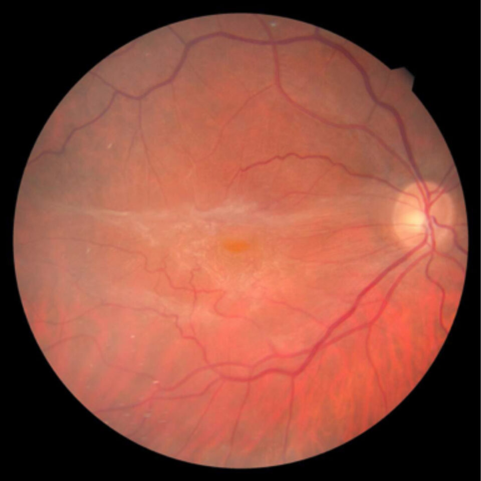
This article examines how clinicians can use OCT images relating to a range of retinal lesions to aid diagnosis and clinical decision making. -
![]() CET module
CET moduleOCT and medical retina: Case Studies
This series of online case studies presents details of the interpretation of OCT images in the diagnosis of a number of retinal conditions, highlighting the distinctive features of each condition and providing pointers on how to avoid misinterpretation. -
![]() CET module
CET moduleAnterior Segment OCT
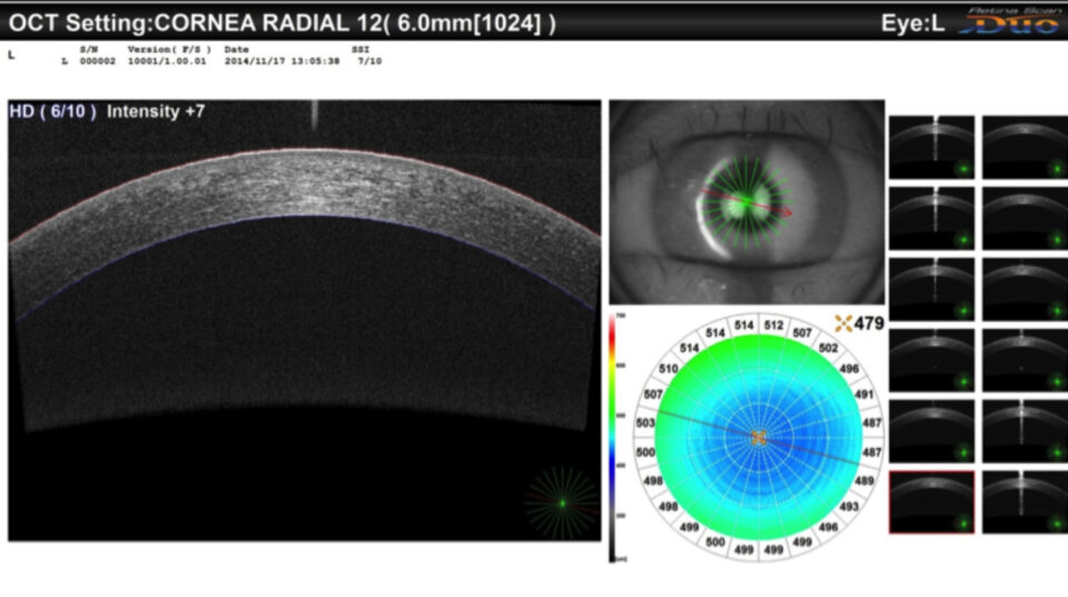
This article examines how practitioners can make effective use of OCT images in the diagnosis and management of a range of anterior chamber structures and conditions. -
![]() CET module
CET moduleOCT in glaucoma: case studies
This series of case studies examines how clinicians can use OCT images to help in diagnosis and referral decision making in glaucoma. -
![]()
Didn’t find what you were looking for?
Check out some of our main topics:

